 Lab Essentials
 AMPIVIEW® RNA probes
 Enabling Your Projects
  GMP Services
 Bulk Solutions
 Research Travel Grant
 Have You Published Using an Enzo Product?
Fodrin, also referred to as non-erythroid (αII-; brain) spectrin, is a tetrameric (αγ)2 actin-binding, fibrous protein, widely distributed in vertebrates, which forms part of the sub-membranous cytoskeleton within many cell types including neurons, and is particularly abundant with axons. The α-subunits of fodrins and spectrin are highly conserved phylogenetically, with the exception of human α-fodrin, which shares only 55-59% homology with erythroid-specific α-spectrins. The β-subunits of spectrin (and γ-subunits of fodrins) are species specific. The interleukin-1 converting enzyme (ICE) family of proteases has been implicated as important effectors of the apoptotic pathway, perhaps acting hierarchically in a protease cascade. Neuronal fodrin is known to be cleaved by calpain following ischaemic insult and it has been proposed that calpain and an unidentified protease play a role in the onset of neuronal death following transient forebrain ischaemia. Recently, an ICE-like protease has been implicated in the early cleavage of fodrin, producing a 150kDa fragment, proximal to CPP32 in Fas-induced and C2-ceramide mediated apoptosis. A cleavage product of α-fodrin has been proposed as a candidate autoantigen in primary Sjögren’s syndrome and α-fodrin has been shown to be the source of a so-called ‘inhibitory protein factor’ family, members of which have been shown to inhibit both GABA and ATP-dependent glutamate uptake into purified synaptic vesicles.
Shipping: Available products typically ship within 24/48h, via priority shipping.
Do you need support? Contact Customer Service or Technical Support.
Online Account
Access or Create Your Account
This antibody is covered by our Worry-Free Guarantee.

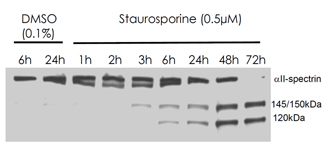
Rat primary septo-hippocampal co-cultures (astrocytes and neurons cultured from day E18 rat embyros) were challenged with the pro-apoptotic compound staurosporine or with vehicle (DMSO) for times indicated above. Protein (40 μg) was separated by SDS-PAGE, transferred to PVDF membrane and probed with FG6090. The caspase-3 cleaved 120-kDa fragment of alpha-II-spectrin was detected at a dilution of 1:4000. Western blot provided by courtesy of Dr Brian Pike, University of Florida.

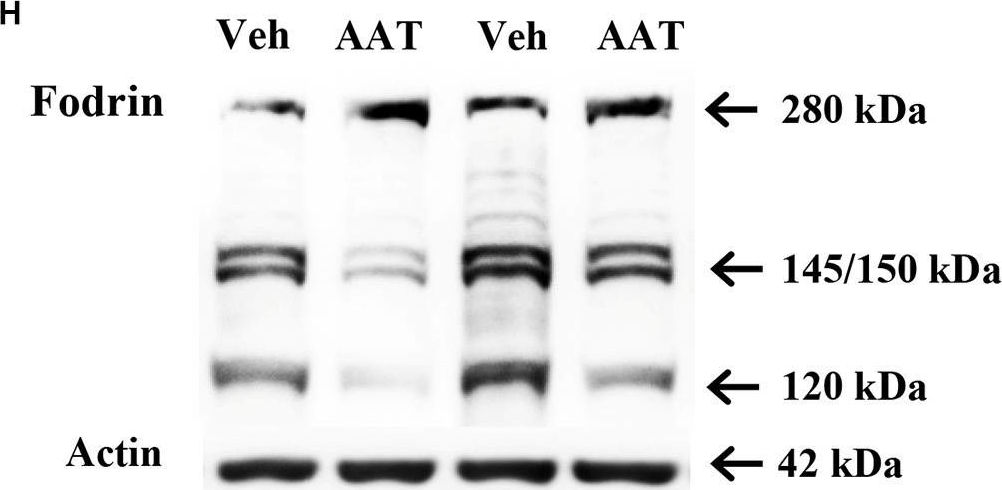
Activation of caspase-3 and calpain in neonatal HI mice after AAT treatment. (A) The photomicrographs show immunochemistry staining of activated caspase-3 in the cortex (Cx), hippocampus (CA1), habenula nucleus (HN), and striatum (Str) at 24 h after HI in the vehicle-treated and AAT-treated groups. (B) Quantification of cleaved caspase-3-labeled cells at 24 h after HI in the Cx, CA1, HN, and Str (n = 14/group, p = 0.005 in Cx, p = 0.437 in CA1, p = 0.006 in HN, p = 0.001 in Str). (C) Quantification of cleaved caspase-3-labeled cells in the cortex at 24 h after HI in male and female mice (n = 7/group, p = 0.008 for males and p = 0.193 for females). (D) Quantification of cleaved caspase-3-labeled cells in the habenula nucleus at 24 h after HI in male and female mice (n = 7/group, p = 0.028 for males and p = 0.067 for females). (E) Quantification of cleaved caspase-3-labeled cells in the striatum at 24 h after HI in male and female mice (n = 7/group, p = 0.059 for males and p = 0.006 for females). (F) The caspase-3 activity in cortical tissue homogenate was measured at 24 h after HI in the AAT-treated and vehicle-treated groups (Veh = 12, AAT = 11, p = 0.011). (G) Measurement of caspase-3 activity at 24 h after HI in male and female mice (Veh = 6, AAT = 5, p = 0.273 in male; n = 6/group, p = 0.015 in female). (H) Representative immunoblotting of fodrin and actin in the cortical tissue from the injured hemisphere of the vehicle and AAT treatment groups at 24 h after HI. (I) Quantification of 120 kDa fragment expression at 24 h after HI in vehicle-treated and AAT-treated groups (n = 12/group, p = 0.024). (J) Quantification of 145/150 kDa fragment expression at 24 h after HI in vehicle-treated and AAT-treated groups (n = 12/group, p = 0.002). (K) Quantification of 120 kDa fragment expression at 24 h after HI in male and female mice (n = 6/group, p = 0.051 for males and p = 0.097 for females). (L) Quantification of 145/150 kDa fragment expression at 24 h after HI in male and female mice (n = 6/group, p = 0.007 for males and p = 0.086 for females). *p < 0.05, **p < 0.01.
Image collected and cropped by CiteAb under a CC-BY license from the following publication: Alpha1-antitrypsin protects the immature mouse brain following hypoxic-ischemic injury. Front Cell Neurosci (2023)

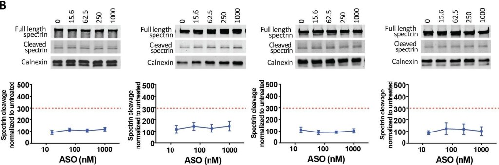
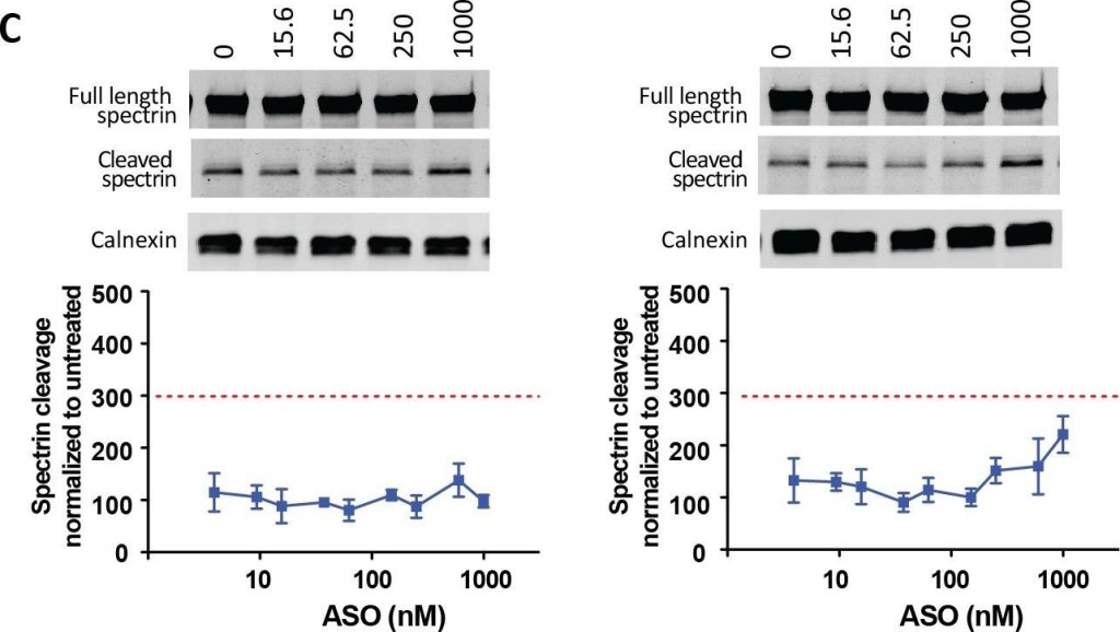
Selection of the best SNP targets.Primary Hu97/18 neurons were treated with 5e-9-5e ASOs targeted to 10 HD-SNPs at 6–1000 nM for 6 days. (A) HTT Western blots and quantitation for the 4 SNPs with the greatest activity. HTT levels are normalized to the internal loading control calnexin and then to the untreated sample for each allele. (B) Western blots showing full length and cleaved spectrin for the 4 ASOs. Spectrin fragment is normalized to calnexin and then to the untreated sample. Membranes were probed for HTT and reprobed for spectrin. Representative images are shown. n = 4–8 per data point. Data are presented as mean ± SD. Two way ANOVA with Bonferroni post hoc test have been performed and p values are illustrated with *, **, ***, **** for p = 0.05, 0.01, 0.001, and 0.0001. The PS backbone is black and MOE modifications are illustrated by orange. The SNP is underlined. The red dashed line represents the toxicity threshold.
Image collected and cropped by CiteAb under a CC-BY license from the following publication: Allele-specific suppression of mutant huntingtin using antisense oligonucleotides: providing a therapeutic option for all Huntington disease patients. PLoS One (2014)

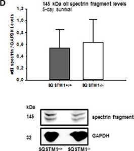
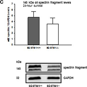
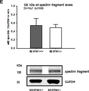
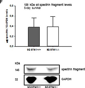
SQSTM1 plays a role in early secondary brain damage formation after TBI. Lesion volume of SQSTM1−/− mice is reduced compared with SQSTM1+/+ littermates 24 h after experimental TBI (A). 5 days after trauma there is no difference between SQSTM1−/− and SQSTM1+/+ mice. Representative cresyl-violet stained sections at the coronal plane from 1.70 mm anterior to bregma, 0.46 mm posterior to bregma, and 1.46 mm posterior to bregma at 24 h (A) and 5 days (B) after CCI are shown (according to The Mouse Brain Library: www.mbl.org). Protein analysis was performed by immunoblotting to determine 120 and 145 kDa αII-spectrin fragments in perilesional brain tissue samples. In SQSTM1−/− mice calpain-dependent spectrin proteolysis to 145/150-kDa fragments is reduced by trend 24 h after CCI (C). There is no effect on calpain-dependent cell death 5 days after CCI (D). SQSTM1−/− does not influence caspase-dependent spectrin proteolysis to 120-kDa fragments (E,F). SQSTM1, sequestosome 1; GAPDH, glyceraldehyde-3-phosphate dehydrogenase, data are expressed as mean ± S.D. *P < 0.05.
Image collected and cropped by CiteAb under a CC-BY license from the following publication: Sequestosome 1 Deficiency Delays, but Does Not Prevent Brain Damage Formation Following Acute Brain Injury in Adult Mice. Front Neurosci (2018)

Targeting two variants of a single HD-SNP to provide a therapeutic option to all HD patients.(A) The genotypes for the sequenced HD population at rs7685686. Green = heterozygous HD population (rs7685686_A/G, 48.7%, targetable by A-series ASOs and rs7685686_G/A, 3.8%, targetable by X-series ASOs). Blue = homozygous HD population (rs7685686_A/A, 44.9%, targetable by A-series ASOs and rs7685686_G/G, 2.6%, targetable by X-series ASOs). Primary YAC128 neurons were treated with ASO at 16–1000 nM for 6 days. (B) Western blot and quantitation of HTT protein levels. HTT levels are normalized to the internal loading control calnexin and then to the untreated sample for each allele. (C) Western blots showing full length and cleaved spectrin. Spectrin fragment is normalized to calnexin and then to the untreated sample. Membranes were probed for HTT and reprobed for spectrin. Representative images are shown. n = 8–12 per data point. Data are presented as mean ± SD. Two way ANOVA with Bonferroni post hoc test have been performed and p values are illustrated with *, **, ***, **** for p = 0.05, 0.01, 0.001, and 0.0001. The PS backbone is represented by black. MOE and cEt modifications are illustrated by orange and blue, respectively. The SNP is underlined. The red dashed line represents the toxicity threshold.
Image collected and cropped by CiteAb under a CC-BY license from the following publication: Allele-specific suppression of mutant huntingtin using antisense oligonucleotides: providing a therapeutic option for all Huntington disease patients. PLoS One (2014)

SQSTM1 plays a role in early secondary brain damage formation after TBI. Lesion volume of SQSTM1−/− mice is reduced compared with SQSTM1+/+ littermates 24 h after experimental TBI (A). 5 days after trauma there is no difference between SQSTM1−/− and SQSTM1+/+ mice. Representative cresyl-violet stained sections at the coronal plane from 1.70 mm anterior to bregma, 0.46 mm posterior to bregma, and 1.46 mm posterior to bregma at 24 h (A) and 5 days (B) after CCI are shown (according to The Mouse Brain Library: www.mbl.org). Protein analysis was performed by immunoblotting to determine 120 and 145 kDa αII-spectrin fragments in perilesional brain tissue samples. In SQSTM1−/− mice calpain-dependent spectrin proteolysis to 145/150-kDa fragments is reduced by trend 24 h after CCI (C). There is no effect on calpain-dependent cell death 5 days after CCI (D). SQSTM1−/− does not influence caspase-dependent spectrin proteolysis to 120-kDa fragments (E,F). SQSTM1, sequestosome 1; GAPDH, glyceraldehyde-3-phosphate dehydrogenase, data are expressed as mean ± S.D. *P < 0.05.
Image collected and cropped by CiteAb under a CC-BY license from the following publication: Sequestosome 1 Deficiency Delays, but Does Not Prevent Brain Damage Formation Following Acute Brain Injury in Adult Mice. Front Neurosci (2018)
| Regulatory Status |
RUO – Research Use Only |
|---|
Last modified: May 29, 2024