The lymphocyte activation gene-3 (LAG-3, CD223), a member of the immunoglobulin superfamily (IgSF) related to CD4, binds to the major histocompatibility complex (MHC) class II molecules but with higher affinity than CD4. Several alternative mRNA splice-variants of human LAG-3 have been described, two of them encoding potential secreted forms: LAG-3V1 (i.e. the D1-D2 domains of the protein, 36 kDa) and LAG-3V3 (D1-D3, 52 kDa). The longer form was detected by ELISA in the serum of healthy individuals as well as of tuberculosis patients with a favorable outcome. LAG-3 expression by T cell clones correlated with IFN-γ production, and hence soluble LAG-3 has been suggested as a serological marker of Th1 responses.
Shipping: Available products typically ship within 24/48h, via priority shipping.
Do you need support? Contact Customer Service or Technical Support.
Online Account
Access or Create Your Account
This antibody is covered by our Worry-Free Guarantee.

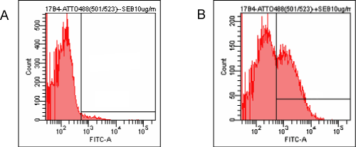
Figure: Detection of endogenous human LAG-3 by FACS analysis using LAG-3 (human), mAb (17B4) (ATTO 488) (Prod. No. ALX-804-806TD). Method: Human PBMC were stimulated (B) or not (A) with 1µg/ml of superantigen SEB. After 2 days, PBMC were stained with 10µg/ml (1µg/0.5×106 cells) of LAG-3 (human), mAb (17B4) (ATTO 488) (Prod. No. ALX-804-806TD) and analyzed by flow cytometry.

Product Details
Alternative Name |
Lymphocyte activation gene-3, FDC protein, CD223 |
---|---|
Application |
Flow Cytometry, ICC |
Clone |
17B4 |
Formulation |
Liquid. In PBS containing 0.1M sodium bicarbonate, 50mM Tris, 1% BSA and 0.02% sodium azide. |
Host |
Mouse |
Immunogen |
Synthetic peptide corresponding to 30 aa (GPPAAAPGHPLAPGPHPAAPSSWGPRPRRY) from the first N-terminal D1 domain of human LAG-3 (lymphocyte activation gene-3). |
Isotype |
IgG1 |
Purity Detail |
Purified from concentrated hybridoma tissue culture supernatant by Protein A Sepharose™ CL-4B Flow Fast Column. |
Recommendation Dilutions/Conditions |
Flow Cytometry (1:100)Suggested dilutions/conditions may not be available for all applications.Optimal conditions must be determined individually for each application. |
Source |
Purified from hybridoma tissue culture supernatant. |
Species Reactivity |
Human |
Specificity |
Recognizes the 30 aa extra-loop of the first N-terminal D1 domain of LAG-3. |
Technical Info / Product Notes |
1 test means: 1µl of MAb is used to stain 500’000 cells in a sample volume of 50µl. ATTO-fluorescent antibodies show increased photostability, outstanding brightness and intense signals. ATTO dyes are thermally stable, resistant to environmental changes and show no significant isomerization. ATTO 488 shows bright green fluorescence (λabs (max): 501nm, λem (max): 523nm, εmax: 90’000). SEPHAROSE is a trademark of GE Healthcare companies. |
UniProt ID |
P18627 |
Worry-free Guarantee |
This antibody is covered by our Worry-Free Guarantee. |
Handling & Storage
Use/Stability |
Stable for at least 6 months after receipt when stored as recommended. |
---|---|
Handling |
Avoid vigorous centrifugation and vortexing. Do not freeze. Protect from light. |
Short Term Storage |
+4°C |
Long Term Storage |
+4°C |
Shipping |
Blue Ice |
Regulatory Status |
RUO – Research Use Only |
---|
- Identification of tonsillar CD4+CD25-LAG3+ T cells as naturally occurring IL-10-producing regulatory T cells in human lymphoid tissue: S. Sumitomo, et al.; J. Autoimmun. 76, 75 (2017), Application(s): Flow cytometry and immunofluorescence, Abstract
- Transcription factor early growth response 3 is associated with the TGF-β1 expression and the regulatory activity of CD4-positive T cells in vivo: Sumitomo, S., Fujio, K., et al.; J. Immunol. 191, 2351 (2013), Abstract
- Human lymphocyte activation gene-3 molecules expressed by activated T cells deliver costimulation signal for dendritic cell activation: C. Casati, et al.; J. Immunol. 180, 3782 (2008), Abstract
- A soluble lymphocyte activation gene-3 (sLAG-3) protein as a prognostic factor in human breast cancer expressing estrogen or progesterone receptors: F. Triebel, et al.; Cancer Lett. 235, 147 (2006), Abstract
- The negative regulatory function of the lymphocyte-activation gene-3 co-receptor (CD223) on human T cells: L. Macon-Lemaitre and F. Triebel; Immunology 115, 170 (2005), Abstract
- Immunological mechanisms elicited at the tumour site by lymphocyte activation gene-3 (LAG-3) versus IL-12: sharing a common Th1 anti-tumour immune pathway: E.D. Carlo, et al.; J. Pathol. 205, 82 (2005), Abstract
- T Lymphocytes infiltrating various tumour types express the MHC class II ligand lymphocyte activation gene-3 (LAG-3): role of LAG-3/MHC class II interactions in cell-cell contacts: C.E. Demeure, et al.; Eur. J. Cancer 37, 1709 (2001), Abstract
- CD3/TCR complex-associated lymphocyte activation gene-3 molecules inhibit CD3/TCR signaling: S. Hannier, et al.; J. Immunol. 161, 4058 (1998), Abstract — Full Text
- Characterization of the major histocompatibility complex class II binding site on LAG-3 protein: B. Huard, et al.; PNAS 94, 5744 (1997), Abstract — Full Text
- Cellular expression and tissue distribution of the human LAG-3-encoded protein, an MHC class II ligand: B. Huard, et al.; Immunogenetics 39, 213 (1994), Abstract
Related Products

Alternative Name | Lymphocyte activation gene-3, FDC protein, CD223 |
---|---|
Application | ICC, IHC, IP, WB |
Host | Mouse |
Isotype | IgG1 |
Species Reactivity | Human |

Alternative Name | Lymphocyte activation gene-3, FDC protein, CD223 |
---|---|
Application | ELISA, WB |
Host | Mouse |
Isotype | IgG1 |
Species Reactivity | Human |

Alternative Name | Lymphocyte activation gene-3, FDC protein, CD223 |
---|---|
Application | Flow Cytometry |
Host | Mouse |
Isotype | IgG1 |
Species Reactivity | Human |

Alternative Name | Lymphocyte activation gene-3, FDC protein, CD223 |
---|---|
Application | Flow Cytometry, ICC |
Host | Mouse |
Isotype | IgG1 |
Species Reactivity | Human |
Datasheet, Manuals, SDS & CofA
Manuals And Inserts
Certificate of Analysis
Please enter the lot number as featured on the product label
SDS
Enzo Life Science provides GHS Compliant SDS
If your language is not available please fill out the SDS request form