 Lab Essentials
 AMPIVIEW® RNA probes
 Enabling Your Projects
  GMP Services
 Bulk Solutions
 Research Travel Grant
 Have You Published Using an Enzo Product?
- Far-red fluorescent specific DNA dye
- Stable and highly pure
- Live, permeable and fixed cells can be analyzed
- No photobleaching effect
- No RNase treatment is required
- GFP and FITC compatible
- UV laser source is not required for excitation
- Validated for a wide range of cell densitites
- Quick and easy to use!
The NUCLEAR-ID® Red DNA Stain is a cell permeable dye, designed for use in a range of fluorescence detection technologies, in the discrimination of nucleated cells. It is resistant to photobleaching and is suitable for live-cell staining of nuclei. Also this dye provides a convenient approach for studying the induction and inhibition of cell cycle progression by flow cytometry. Potential applications of this reagent for live-cell studies are in the determination of cellular DNA content and cell cycle distribution, for the detection of variations in growth patterns, for monitoring apoptosis, and for evaluating tumor cell behavior and suppressor gene mechanisms.
Shipping: Available products typically ship within 24/48h, via priority shipping.
Do you need support? Contact Customer Service or Technical Support.
Online Account
Access or Create Your Account
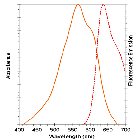
NUCLEAR-ID® Red DNA stain spectra

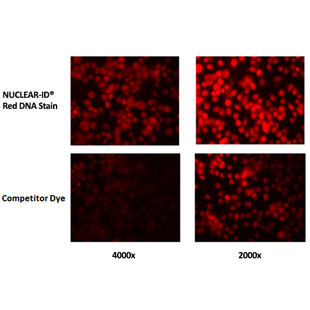
NUCLEAR-ID Red DNA Stain requires lower concentration than competitor’s dye to visualize dsDNA.HeLa cells were grown to ~60% confluency. Cells were stained with NUCLEAR-ID Red DNA Stain at a final concentration of 4000x or 2000x or a competitor’s dye at the equivalent µM concentration at 37°C and gently washed post-staining. Cells were imaged at 15 min.
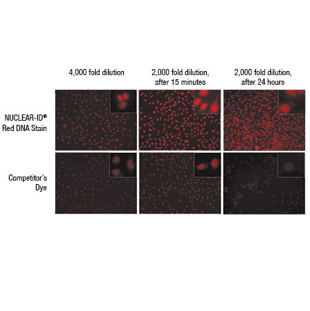
NUCLEAR-ID Red DNA Stain requires lower concentration than competitor’s dye to visualize dsDNA.HeLa cells were grown to ~60% confluency. Cells were stained with NUCLEAR-ID Red DNA Stain at a final concentration of 4000x or 2000x or with a competitor’s dye at an equivalent µM concentration at 37°C and gently washed post-staining. Cells were imaged at 15 min and 24h. Results show that 4000x NUCLEAR-ID Red DNA Stain was required for visualization of the dsDNA, while equivalent to 2000x was required for the competitor’s dye. At 24h, the competitor’s dye intensity and cell growth were dramatically reduced at the 2000x equivalent final concentration. At the same time point, 2000x of NUCLEAR-ID Red DNA Stain did not affect cell growth or fluorescent intensity. The NUCLEAR-ID Red DNA Stain shows lower cytotoxicity and requires lower concentration in live cell studies, resulting in lower costs.
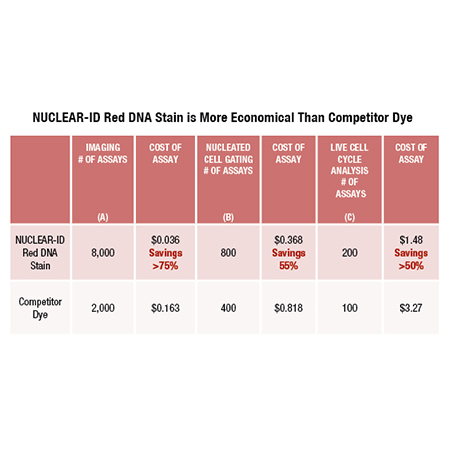
Relative costs of using NUCLEAR-ID Red DNA in comparison to competitor dye in various applications: (A) Imaging (visualization), (B) Nucleated Cell Gating (flow cytometry) and (C) Live Cell Cycle analysis using flow cytometry. Dilutions can vary depending on cell strain and cell concentration.Notes:Assumes staining of a 100 µL staining volumeAssumes staining of a 500 µL cell suspension volumeAssumes a staining of 500 µL cell suspension volume


| Regulatory Status |
RUO – Research Use Only |
|---|
Related Products
NUCLEAR-ID® Red cell cycle kit (GFP-CERTIFIED®)
ENZ-51008
Convenient kit for studying cell cycle progression by various applications
| Application | Flow Cytometry, Fluorescence microscopy |
|---|
NUCLEAR-ID® Green cell cycle kit
ENZ-51014
Cell Cycle Results Independent of Incubation Time, Temperature, Dye and Cell Concentrations
| Application | Flow Cytometry, Fluorescence microscopy |
|---|
Last modified: May 29, 2024