Photostable, non-toxic and selective mitochondrial dye that stains regardless of membrane potential
- Mitochondria-selective dye stains live, detergent permeabilized and aldehyde fixed cells
- Long wavelength red emission is easily multiplexed with common fluorescent dyes
- Highly resistant to photobleaching and concentration quenching, for strong, consistent fluorescent signal
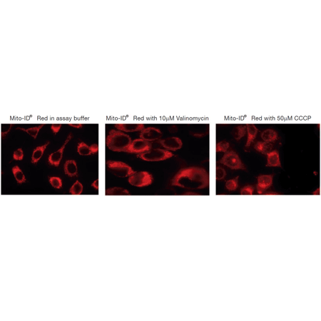
- Highlights mitochondria regardless of the organelle’s membrane potential status
- Stringently manufactured, to control and eliminate non-specific assay artifacts
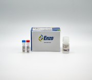
MITO-ID® Red Detection Kit (GFP-CERTIFIED®) contains a proprietary membrane-permeable mitochondria-selective dye suitable for use with live-, detergent-permeabilized- and even aldehyde-fixed-cells. Unlike conventional dyes, such as DiOC6(3), JC-1, rhodamine 123 and tetramethylrhodamine ethyl ester, MITO-ID® Red dye highlights mitochondria regardless of their energetic state. The dye is compatible with most fluorescence detection systems, including conventional and confocal fluorescence microscopes, as well as, High Content Screening (HCS) platforms. The kit is useful for assessing mitochondrial morphology changes, estimating mitochondrial mass and co-localizing GFP-tagged proteins to the mitochondrial compartment. This kit is specifically designed for use with GFP expressing cell lines, as well as cells expressing blue, cyan or yellow fluorescent proteins (BFPs, CFPs, YFPs). Additionally, the kit is suitable for use with live or post-fixed cells in conjunction with fluorescent probes, such as labeled antibodies, or other fluorescent conjugates displaying similar spectral properties such as fluorescein and coumarin. A nuclear counterstain (Hoechst 33342) is provided to highlight this organelle as well. Wavelength maxima: MITO-ID® Red λex 558 nm, λem 690 nm; Hoechst 33342 λex 350 nm, λem 461 nm.
Shipping: Available products typically ship within 24/48h, via priority shipping.
Do you need support? Contact Customer Service or Technical Support.
Online Account
Access or Create Your Account
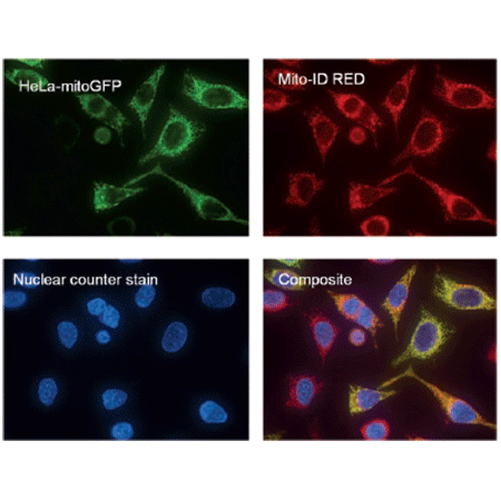
HeLa-TurboGreen-mitochondria cells (HeLa-mitoGFP, MarinPharm GmbH, Luckenwalde, Germany) stained with MITO-ID® Red and Hoechst 33342 (blue) dyes. MITO-ID® Red co-localizes with the EGFP-cytochrome C oxidase signal (yellow signal), demonstrating selectivity for mitochondria. Note that mitochondria in cells no longer expressing the GFP-tagged protein appear red in the composite image.
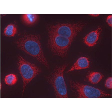
Composite fluorescence microscopy images of HeLa cells (40X objective lens). Cells were stained with MITO-ID® Red dye for 15 minutes. Nuclei were counter-stained with Hoechst 33342 dye.


Product Details
| Application |
Fluorescence microscopy, Fluorescent detection |
|---|---|
| Application Notes |
For use with GFP-expressing cell lines, as well as cells expressing blue, cyan or yellow fluorescent proteins. |
| Contents |
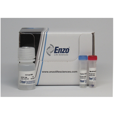
MITO-ID® Red Detection Reagent |
| Quality Control |
A sample from each lot of GFP-CERTIFIED® MITO-ID® Red Mitochondrial Detection Kit is used to stain HeLa cells, expressing GFP-cytochrome C oxidase, using the procedures described in the user manual. The selectivity of the MITO-ID® Red dye is evident as shown by the co-localization with a GFP-cytochrome C oxidase fusion expressed in the HeLa cells. |
| Quantity |
For ENZ-51007-500, 500 assays |
| Technical Info / Product Notes |
Application Note: |
Handling & Storage
| Use/Stability |
With proper storage, the kit components are stable up to the date noted on the product label. Store kit at -20°C in a non-frost free freezer, or –80°C for longer term storage. |
|---|---|
| Handling |
Protect from light. Avoid freeze/thaw cycles. |
| Long Term Storage |
-80°C |
| Shipping |
Dry Ice |
| Regulatory Status |
RUO – Research Use Only |
|---|
- The Changes in Mitochondrial Morphology and Physiology Accompanying Apoptosis in Galleria mellonella (Lepidoptera) Immunocompetent Cells during Conidiobolus coronatus (Entomophthorales) Infection: A. Kaczmarek, et al.; Int. J. Mol. Sci. 24, 10169 (2023), Abstract
- Human anti-smallpox long-lived memory B cells are defined by dynamic interactions in the splenic niche and long-lasting germinal center imprinting: Chappert, P., Huetz, F., et al.; Immunity 55, 1872 (2022), Abstract
- Subcellular Redox Responses Reveal Different Cu-dependent Antioxidant Defenses between Mitochondria and Cytosol: Zhang, Y., Wen, M., et al.; bioRxiv , (2022)
- Intracellular reactive oxygen species trafficking participates in seed dormancy alleviation in Arabidopsis seeds: R. Jurdak, et al.; New Phytol. , (2022), Abstract
- Unveiling luminescent IrI and RhI N‐heterocyclic carbene complexes: structure, photophysical specifics, and cellular localization in the endoplasmic reticulum: I.M. Daubit, et al.; Chemistry 27, 6783 (2021), Application(s): Fluorescence microscopy, Abstract — Full Text
- Intercellular Mitochondria Transfer to Macrophages Regulates White Adipose Tissue Homeostasis and Is Impaired in Obesity: Brestoff, J. R., Wilen, C. B., et al.; Cell Metab. 33, 270 (2021), Abstract
- Suppression of optineurin impairs the progression of hepatocellular carcinoma through regulating mitophagy: S. Inokuchi, et al.; Cancer Med. 10, 1501 (2021), Application(s): Fluorescence microscopy, Abstract — Full Text
- Retrograde signalling from the mitochondria to the nucleus translates the positive effect of ethylene on dormancy breaking of Arabidopsis thaliana seeds: R. Jurdak, et al.; New Phytol. 229, 2192 (2021), Application(s): Fluorescence microscopy on seeds, Abstract
- Progesterone receptor membrane associated component 1 enhances obesity progression in mice by facilitating lipid accumulation in adipocytes: R. Furuhata, et al.; Commun. Biol. 3, 479 (2020), Abstract — Full Text
- Perfusion reduces bispecific antibody aggregation via mitigating mitochondrial dysfunction-induced glutathione oxidation and ER stress in CHO cells: P. Sinharoy, et al.; Sci. Rep. 10, 16620 (2020), Abstract — Full Text
- Mitochondrial 4-HNE derived from MAO-A promotes mitoCa2+ overload in chronic postischemic cardiac remodeling: Y. Santin, et al.; Cell Death Differ. 27, 1907 (2020), Application(s): Fluorescence microscopy, Abstract — Full Text
- CXCR2 specific endocytosis of immunomodulatory peptide LL-37 in human monocytes and formation of LL-37 positive large vesicles in differentiated: Z. Zhang, et al.; Bone Rep. 12, 100237 (2019), Application(s): Fluorescence microscopy, Abstract — Full Text
- Heat shock factor 1 is a direct antagonist of AMP-activated protein kinase: K.H. Su, et al.; Mol. Cell 76, 546 (2019), Application(s): Flow cytometry, Abstract — Full Text
- Mitochondrial cysteinyl-tRNA synthetase is expressed via alternative transcriptional initiation regulated by energy metabolism in yeast cells: A. Nishimura, et al.; J. Biol. Chem. 294, 13781 (2019), Application(s): Fluorescence microscopy on yeasts, Abstract — Full Text
- The bovine herpesvirus-1 major tegument protein, VP8, interacts with host HSP60 concomitant with deregulation of mitochondrial function: S. Afroz, et al.; Virus Res. 261, 37 (2019), Application(s): Fluorescence microscopy, Abstract
- Mitochondrial membrane potential identifies cells with high recombinant protein productivity: L. Chakrabarti, et al.; J. Immunol. Methods 464, 31 (2019), Application(s): Cell sorting by FACS, Abstract
- Application of Imaging Flow Cytometry for the Characterization of Intracellular Attributes in Chinese Hamster Ovary Cell Lines at the Single-Cell Level: E. Pekle, et al.; Biotechnol. J. 14, e1800675 (2019), Application(s): High-throughput analysis of CHO cells with flow cytometry, Abstract
- Role of arginase 2 in systemic metabolic activity and adipose tissue fatty acid metabolism in diet-induced obese mice: R.T. Atawia, et al.; Int. J. Mol. Sci. 20, 1462 (2019), Application(s): Fluorescence microscopy using tissue sections, Abstract — Full Text
- Overexpression of TFF3 is involved in prostate carcinogenesis via blocking mitochondria-mediated apoptosis: J. Liu, et al.; Exp. Mol. Med. 50, 110 (2018), Abstract — Full Text
- CD4sup>+/sup> T cell fate decisions are stochastic, precede cell division, depend on GITR co-stimulation, and are associated with uropodium development: Cobbold, S. P., Adams, E., et al.; bioRxiv , (2018)
- Temozolomide-induced increase of tumorigenicity can be diminished by targeting of mitochondria in in vitro models of patient individual glioblastoma: D. William, et al.; PLoS One. 13, e0191511 (2018), Application(s): Fluorescence microscopy, Abstract
- CD4+ T cell fate decisions are stochastic, precede cell division, depend on GITR co-stimulation, and are associated with uropodium development: S.P. Cobbold, et al.; Front. Immunol. 9, 1381 (2018), Application(s): Fluorescence microscopy / Reactant(s): Mouse, Abstract — Full Text
- Long-circulating amphiphilic doxorubicin for tumor mitochondria-specific targeting: J. Xi, et al.; ACS Appl. Mater. Interfaces 10, 43482 (2018), Application(s): Fluorescence microscopy on tissue section, Abstract — Full Text
- Functional characterization of the first filamentous fungal tRNA-isopentenyltransferase and its role in the virulence of Claviceps purpurea: J. Hinsch, et al.; New Phytol. 211, 980 (2016), Abstract
- Mitochondrial targeting increases specific activity of a heterologous valine assimilation pathway in Saccharomyces cerevisiae: K.V. Solomon, et al.; Metab. Eng. Commun. 3, 68 (2016), Application(s): Protein localization, Abstract — Full Text
- Effects of two fullerene derivatives on monocytes and macrophages: S. Pacor, et al.; Biomed. Res. Int. 2015, Article ID 915130 (2015), Application(s): Confocal microscopy on monocytes and macrophages, Abstract — Full Text
- Induction of androgen formation in the male by a TAT-VDAC1 fusion peptide blocking 14-3-3ɛ protein adaptor and mitochondrial VDAC1 interactions: Y. Aghazadeh, et al.; Mol. Ther. 22, 1779 (2014), Abstract
- Protein modifications regulate the role of 14-3-3γ adaptor protein in cAMP-induced steroidogenesis in MA-10 Leydig cells: Y. Aghazadeh, et al.; J. Biol. Chem. 289, 26542 (2014), Abstract
- Mitochondrial fission induced by platelet-derived growth factor regulates vascular smooth muscle cell bioenergetics and cell proliferation: J.K. Salabei, et al.; Redox Biol. 1, 542 (2013), Application(s): Detection by confocal microscopy, Abstract — Full Text
- Utilization of fluorescent probes for the quantification and identification of subcellular proteomes and biological processes regulated by lipid peroxidation products: T.D. Cummins, et al.; Free Radic. Biol. Med. 59, 56 (2013), Application(s): Detection by confocal microscopy, Abstract — Full Text
- Ectopic ATP synthase blockade suppresses lung adenocarcinoma growth by activating the unfolded protein response: H.Y. Chang, et al.; Cancer Res. 72, 4696 (2012), Abstract — Full Text
- Hormone-induced 14-3-3γ adaptor protein regulates steroidogenic acute regulatory protein activity and steroid biosynthesis in MA-10 Leydig cells: Y. Aghazadeh, et al.; J. Biol. Chem. 287, 15380 (2012), Application(s): Detection of mitochondria in MA-10 mouse Leydig tumor cells using confocal microscopy, Abstract — Full Text
- Intracellular Energetic Units regulate metabolism in cardiac cells: V. Saks, et al.; J. Mol. Cell. Cardiol. 52, 419 (2012), Application(s): Detection of mitochondria in cardiomyocytes using confocal microscopy, Abstract
- High-mobility group A1 protein inhibits p53-mediated intrinsic apoptosis by interacting with Bcl-2 at mitochondria: F. Esposito, et.al; Cell Death Dis. 3, e383 (2012), Application(s): Mitochondrial staining in HEK293cells transfected with EGFP-HMGA1b, Abstract — Full Text
- Opa3, a novel regulator of mitochondrial function, controls thermogenesis and abdominal fat mass in a mouse model for Costeff syndrome: T. Wells, et al.; Hum. Mol. Genet. 18, 4836 (2012), Application(s): Visualization of mitochondria in paraffin embedded sections of mouse brown adipose tissue with Mito-ID® Red detection kit., Abstract — Full Text
- Cytometric assessment of mitochondria using fluorescent probes: C. Cottet-Rousselle, et al.; Cytometry A 79, 405 (2011), Abstract — Full Text
- The targeting of plasmalemmal ceramide to mitochondria during apoptosis: E.B. Babiychuk, et al.; PLoS One 6, e23706 (2011), Application(s): Detection of mitochondria in T cell and monocyte cell lines using fluorescence microscopy, Abstract — Full Text
- Mitochondria-cytoskeleton interaction: distribution of β-tubulins in cardiomyocytes and HL-1 cells: R. Guzun, et al.; Biochim. Biophys. Acta 1807, 458 (2011), Application(s): Detection of mitochondria in cardiomyocytes using fluorescence microscopy, Abstract
Related Products
LYSO-ID® Red detection kit (GFP-CERTIFIED®)
ENZ-51005
Acidic organelle-selective dye for live cell staining of lysosomes

| Application | Fluorescence microscopy, Fluorescent detection |
|---|
MITO-ID® Green detection kit
ENZ-51022
Photostable, non-toxic and selective mitochondrial dye that stains regardless of membrane potential

| Application | Fluorescence microscopy, Fluorescent detection |
|---|

| Application | Fluorescence microscopy |
|---|
ER-ID® Red assay kit (GFP-CERTIFIED®)
ENZ-51026
Widely cited Endoplasmic Reticulum staining with minimal toxicity to cells
| Application | Fluorescence microscopy |
|---|
Last modified: May 29, 2024
Datasheet, Manuals, SDS & CofA
Certificate of Analysis
Please enter the lot number as featured on the product label
SDS
Enzo Life Science provides GHS Compliant SDS
If your language is not available please fill out the SDS request form
 Lab Essentials
 AMPIVIEW® RNA probes
 Enabling Your Projects
  GMP Services
 Bulk Solutions
 Research Travel Grant
 Have You Published Using an Enzo Product?
