 Lab Essentials
 AMPIVIEW® RNA probes
 Enabling Your Projects
  GMP Services
 Bulk Solutions
 Research Travel Grant
 Have You Published Using an Enzo Product?
- Dual-emission dye fluoresces either green or orange depending upon mitochondrial membrane potential status
- 10X more sensitive than JC-1 with superior aqueous solubility
- True mix-and-read homogeneous assay for live cells
- Suitable for chemical/environmental toxicity screening
- Optimized for fluorescence microscopy, flow cytometry and microplate reader
- Suitable for high-throughput applications
Enzo Life Sciences MITO-ID® Membrane Potential Detection Kit is a mitochondria tracker dye including a dual-emission cationic dye. It measures the mitochondrial membrane potential (MMP) in live cells. In energized or active cells, the MITO-ID® Membrane Potential dye rapidly accumulates as orange-fluorescent aggregates in the mitochondria due to their relative negative charge, while it exists as a green-fluorescent monomer in the cytosol. However, in cells with compromised MMP, the MITO-ID® Membrane Potential dye exists primarily as green-fluorescent monomers throughout the cytosol and no longer exhibits orange fluorescence in the mitochondria. A widely used uncoupler of mitochondrial oxidative phosphorylation (CCCP) is provided as a positive control for monitoring loss in mitochondrial membrane potential.
Cell-based assays for evaluating the functional status of mitochondria are useful tools for elucidating the role of mitochondrial activity in drug-induced toxicity, the apoptosis cascade and other cellular and biochemical processes. The loss of the mitochondrial membrane potential (MMP) is often associated with early stages of apoptosis. The collapse of MMP coincides with the opening of the mitochondrial permeability transition pores, leading to the release of cytochrome C into the cytosol, which in turn triggers other downstream events in the apoptotic cascade.
Shipping: Available products typically ship within 24/48h, via priority shipping.
Do you need support? Contact Customer Service or Technical Support.
Online Account
Access or Create Your Account
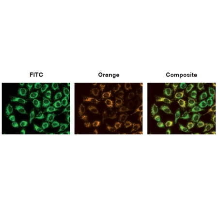
Figure 1: The mitochondria of HeLa cells were stained with MITO-ID® Membrane Potential reagent, and visualized by epifluorescence microscopy. Orange fluorescent aggregates are localized in the mitochondria (Orange channel), while green fluorescent monomers mainly stain the cytosol (FITC channel).
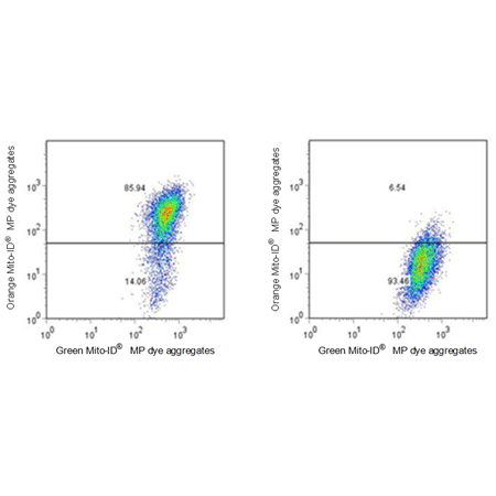
Figure 2: Flow Cytometric Analysis of Control and Treated Cells. Jurkat cells were untreated (left) and were treated with 1 μM CCCP for 15 mins (right). Cells were then stained with Enzo MITO-ID® Membrane Potential Dye, and run on a FACS Calibur instrument.


| Regulatory Status |
RUO – Research Use Only |
|---|
Related Products
MITO-ID® Membrane potential cytotoxicity kit
ENZ-51019
A Real-time Mitochondrial membrane Potential Assay with Superior Sensitivity

| Application | HTS, Microplate |
|---|
Last modified: May 29, 2024