The lymphocyte activation gene-3 (LAG-3, CD223), a member of the immunoglobulin superfamily (IgSF) related to CD4, binds to the major histocompatibility complex (MHC) class II molecules but with higher affinity than CD4. Several alternative mRNA splice-variants of human LAG-3 have been described, two of them encoding potential secreted forms: LAG-3V1 (i.e. the D1-D2 domains of the protein, 36 kDa) and LAG-3V3 (D1-D3, 52 kDa). The longer form was detected by ELISA in the serum of healthy individuals as well as of tuberculosis patients with a favorable outcome. LAG-3 expression by T cell clones correlated with IFN-γ production, and hence soluble LAG-3 has been suggested as a serological marker of Th1 responses.
Shipping: Available products typically ship within 24/48h, via priority shipping.
Do you need support? Contact Customer Service or Technical Support.
Online Account
Access or Create Your Account
This antibody is covered by our Worry-Free Guarantee.

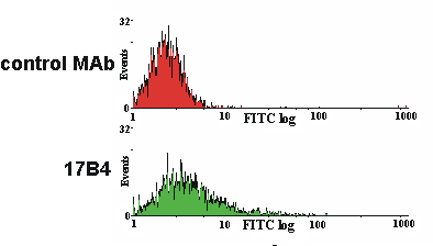
Figure: LAG-3 expression on activated human peripheral blood mononuclear cells (PBMC) detected with LAG-3 (human), mAb (17B4) (FITC) (Prod. No. ALX-804-806F).Method: T lymphocytes from human PBMC are stimulated with 1µg/ml of PHA for three days. Then, after seven days of culture, 3×10⁶ three-days PHA-activated human PBMC are treated with LAG-3 (human), mAb (17B4) (FITC) (Prod. No. ALX-804-806F) or FITC coupled isotype-matched (IgG1) control MAb (used at a saturating dilution of 1/800 and 1/150 respectively) for 30 min. at 4°C in RPMI 1640 and washed twice with 1x PBS. Stained cells are then analysed by FACS [4].

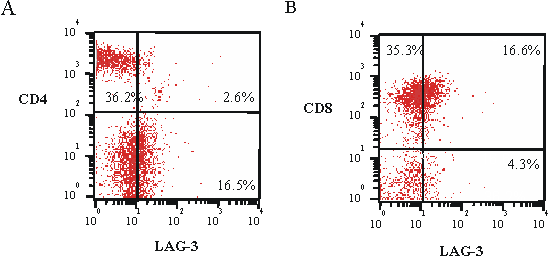
Figure: Expression of LAG-3 on CD4+ and CD8+ subpopulations of tumour infiltrating lymphocytes (TILs) detected with LAG-3 (human), mAb (17B4) (FITC) (Prod. No. ALX-804-806F).Method: TILs from a dissociated renal cell carcinoma sample, stained with 5µg/ml LAG-3 (human), mAb (17B4) (FITC) (Prod. No. ALX-804-806F) and FITC-coupled anti-CD4 or -CD8, are analyzed by a two-colour FACS analysis. Additional staining with anti-CD3 allowed a gate analysis of total T cells. Values indicate percentages in each quadrant [5].

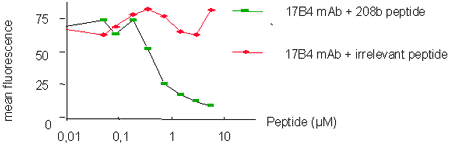
Figure: Specific inhibition of 17B4 staining.Method: LAG-3 (human), mAb (17B4) (FITC) (Prod. No. ALX-804-806F) (10µg/ml) (is preincubated with a specific peptide epitope (208b) or a control tetanus toxoid (TT) peptide at different molarities prior to staining of TILs. Stained cells are then analyzed by FACS.

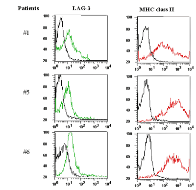
Figure: Tumor infiltrating lymphocytes (TILs) express LAG-3 (detected using LAG-3 (human), mAb (17B4) (Prod. No. ALX-804-806)). Method: Freshly dissociated single cell suspensions of renal cell carcinoma TILs are incubated with LAG-3 (human), mAb (17B4) (FITC) (Prod. No. ALX-804-806) (5µg/ml) and anti-MHC Class II molecules (PE) for 30 min. and washed twice in saline buffer. Additional staining with anti-CD3 allowed a gate analysis of total T cells. The LAG-3 and MHC II profiles of CD3+-gated cells for 3 patients are shown [5].


Product Details
Alternative Name |
Lymphocyte activation gene-3, FDC protein, CD223 |
---|---|
Application |
Flow Cytometry |
Application Notes |
Excellent for Flow Cytometry. |
Clone |
17B4 |
Formulation |
Liquid. In PBS containing 50mM Tris-HCl, 1% BSA and 0.02% sodium azide. |
Host |
Mouse |
Immunogen |
Synthetic peptide corresponding to 30 aa (GPPAAAPGHPLAPGPHPAAPSSWGPRPRRY) from the first N-terminal D1 domain of human LAG-3 (lymphocyte activation gene-3). |
Isotype |
IgG1 |
Purity Detail |
Purified from concentrated hybridoma tissue culture supernatant by Protein A Sepharose™ CL-4B Flow Fast Column. |
Source |
Purified from hybridoma tissue culture supernatant. |
Species Reactivity |
Human |
Specificity |
Recognizes the 30 aa extra-loop of the first N-terminal D1 domain of LAG-3. |
Technical Info / Product Notes |
SEPHAROSE is a trademark of GE Healthcare companies. |
UniProt ID |
P18627 |
Worry-free Guarantee |
This antibody is covered by our Worry-Free Guarantee. |
Handling & Storage
Use/Stability |
Stable for at least 6 months after receipt when stored as recommended. |
---|---|
Handling |
Do not freeze. Protect from light. |
Short Term Storage |
+4°C |
Long Term Storage |
+4°C |
Shipping |
Blue Ice |
Regulatory Status |
RUO – Research Use Only |
---|
- Preoperative immune checkpoint inhibition and cryoablation in early-stage breast cancer: Comen, E., Budhu, S., et al.; iScience 27, 108880 (2024), Abstract
- Nivolumab plus ipilimumab in advanced salivary gland cancer: a phase 2 trial: Vos, J., Burman, B., et al.; Nat. Med. 29, 3077 (2023), Abstract
- Brain radiotherapy, tremelimumab-mediated CTLA-4-directed blockade +/− trastuzumab in patients with breast cancer brain metastases: D.B. Page, et al.; NPJ Breast Cancer 8, 50 (2022), Abstract
- Immunological status of peripheral blood is associated with prognosis in patients with bone and soft-tissue sarcoma: Kim, Y., Kobayashi, E., et al.; Oncol. Lett. 21, 212 (2021), Abstract
- Analysis of CAR-Mediated Tonic Signaling: H. Calderon, et al.; Methods Mol. Biol. 2086, 223 (2020), Abstract
- Targeting VEGFR2 with Ramucirumab strongly impacts effector/ activated regulatory T cells and CD8+ T cells in the tumor microenvironment: Tada, Y., Togashi, Y., et al.; J. Immunother. Cancer 6, 106 (2018), Abstract
- Improving CART-Cell Therapy of Solid Tumors with Oncolytic Virus-Driven Production of a Bispecific T-cell Engager: A. Wing, et al.; Cancer Immunol. Res. 6, 605 (2018), Application(s): Flow Cytometry, Abstract
- NK cell and IFN signatures are positive prognostic biomarkers for resectable pancreatic cancer: M. Hoshikawa, et al.; Biochem. Biophys. Res. Commun. 495, 2058 (2018), Application(s): Flow cytometry, Abstract
- PEGylated IL-10 (Pegilodecakin) Induces Systemic Immune Activation, CD8+ T Cell Invigoration and Polyclonal T Cell Expansion in Cancer Patients: A. Naing, et al.; Cancer Cell 34, 775 (2018), Abstract
- Tumor-infiltrating and peripheral blood T cell immunophenotypes predict early relapse in localized clear cell renal cell carcinoma: N.A. Giraldo, et al.; Clin. Cancer Res. 23, 4416 (2017), Application(s): Flow cytometry, Abstract
- CD26-Mediated Induction of EGR2 and IL-10 as Potential Regulatory Mechanism for CD26 Costimulatory Pathway: R. Hatano, et al.; J. Immunol. 194, 960 (2015), Abstract
- Elevated expression of LAG-3, but not PD-1, is associated with impaired iNKT cytokine production during chronic HIV-1 infection and treatment: J.A. Juno, et al.; Retrovirology 12, 17 (2015), Application(s): Flow Cytometry, Abstract — Full Text
- Enrichment of LAG-3, but not PD-1, on Double Negative T Cells at the Female Genital Tract: J.A. Juno, et al.; Am. J. Reprod. Immunol. 72, 534 (2014), Abstract
- Fusion of Ubiquitin to HIV Gag Impairs Human Monocyte-Derived Dendritic Cell Maturation and Reduces Ability to Induce Gag T Cell Responses: S. Herath, et al.; PLoS One 9, e88327 (2014), Application(s): Flow cytometry using human T-cells, Abstract — Full Text
- PD-1 identifies the patient-specific CD8+ tumor-reactive repertoire infiltrating human tumors: A. Gros, et al.; J. Clin. Invest. 124, 2246 (2014), Application(s): Flow cytometry using human peripheral blood lymphocytes (PBL) and metastatic melanoma cells, Abstract — Full Text
- Selection of CD8+PD-1+ lymphocytes in fresh human melanomas enriches for tumor-reactive T cells: T. Inozume, et al.; J. Immunother. 33, 956 (2010), Application(s): FC analysis with human tumor digests or cultured tumor-infiltrating lymphocytes, Abstract — Full Text
- Human lymphocyte activation gene-3 molecules expressed by activated T cells deliver costimulation signal for dendritic cell activation: C. Casati, et al.; J. Immunol. 180, 3782 (2008), Abstract
- Human dendritic cells acquire a semimature phenotype and lymph node homing potential through interaction with CD4+CD25+ regulatory T cells: J.Bayry, et al.; J. Immunol. 178, 4184 (2007), Application(s): FC analysis with human regulatory T cells, Abstract — Full Text
- A soluble lymphocyte activation gene-3 (sLAG-3) protein as a prognostic factor in human breast cancer expressing estrogen or progesterone receptors: F. Triebel, et al.; Cancer Lett. 235, 147 (2006), Abstract
- Immunological mechanisms elicited at the tumour site by lymphocyte activation gene-3 (LAG-3) versus IL-12: sharing a common Th1 anti-tumour immune pathway: E.D. Carlo, et al.; J. Pathol. 205, 82 (2005), Abstract
- The negative regulatory function of the lymphocyte-activation gene-3 co-receptor (CD223) on human T cells: L. Macon-Lemaitre and F. Triebel; Immunology 115, 170 (2005), Abstract
- Responses to human CD40 ligand/human interleukin-2 autologous cell vaccine in patients with B-cell chronic lymphocytic leukemia: E. Biagi, et al.; Clin. Cancer Res. 11, 6916 (2005), Abstract — Full Text
- T Lymphocytes infiltrating various tumour types express the MHC class II ligand lymphocyte activation gene-3 (LAG-3): role of LAG-3/MHC class II interactions in cell-cell contacts: C.E. Demeure, et al.; Eur. J. Cancer 37, 1709 (2001), Abstract
- CD3/TCR complex-associated lymphocyte activation gene-3 molecules inhibit CD3/TCR signaling: S. Hannier, et al.; J. Immunol. 161, 4058 (1998), Abstract — Full Text
- Characterization of the major histocompatibility complex class II binding site on LAG-3 protein: B. Huard, et al.; PNAS 94, 5744 (1997), Abstract — Full Text
- Cellular expression and tissue distribution of the human LAG-3-encoded protein, an MHC class II ligand: B. Huard, et al.; Immunogenetics 39, 213 (1994), Abstract
- Characterization of the lymphocyte activation gene 3-encoded protein. A new ligand for human leukocyte antigen class II antigens: E. Baixeras, et al.; J. Exp. Med. 176, 327 (1992), Abstract — Full Text
Related Products

Alternative Name | Lymphocyte activation gene-3, FDC protein, CD223 |
---|---|
Application | ICC, IHC, IP, WB |
Host | Mouse |
Isotype | IgG1 |
Species Reactivity | Human |

Alternative Name | Lymphocyte activation gene-3, FDC protein, CD223 |
---|---|
Application | ELISA, WB |
Host | Mouse |
Isotype | IgG1 |
Species Reactivity | Human |

Alternative Name | Lymphocyte activation gene-3, FDC protein, CD223 |
---|---|
Application | Flow Cytometry, ICC |
Host | Mouse |
Isotype | IgG1 |
Species Reactivity | Human |

Alternative Name | Lymphocyte activation gene-3, FDC protein, CD223 |
---|---|
Application | Flow Cytometry, ICC |
Host | Mouse |
Isotype | IgG1 |
Species Reactivity | Human |
Datasheet, Manuals, SDS & CofA
Manuals And Inserts
Certificate of Analysis
Please enter the lot number as featured on the product label
SDS
Enzo Life Science provides GHS Compliant SDS
If your language is not available please fill out the SDS request form