Mitochondria dye
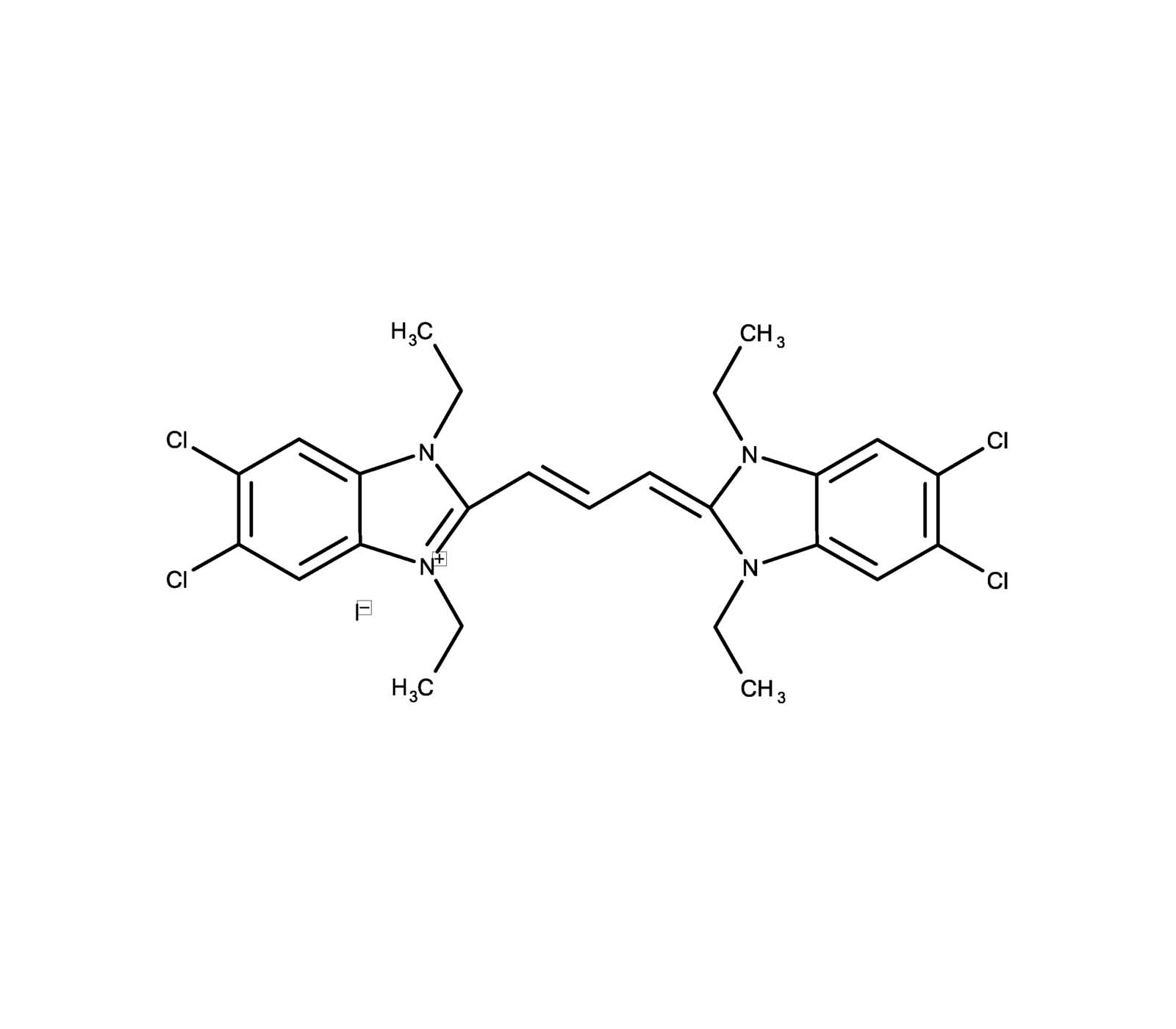
JC-1 is widely used for determining mitochondrial membrane potential by flow cytometry, fluorescence microscopy and in microplate-based fluorescent assays. JC-1 accumulates in mitochondria, selectively generating an orange J-aggregate emission profile (590 nm) in healthy cells. However, upon cell injury, as membrane potential decreases, JC-1 monomers are generated, resulting in a shift to green emission (529 nm). The principal advantage of JC-1 relative to other commonly employed fluorescent probes of mitochondrial membrane potential is that it allows for both qualitative visualization, considering the shift from orange to green fluorescence emission, and quantitative detection, considering the fluorescence intensity ratio. Wavelength Maxima: Excitation 515nm, Emission 529nm
Shipping: Available products typically ship within 24/48h, via priority shipping.
Do you need support? Contact Customer Service or Technical Support.
Online Account
Access or Create Your Account
Product Details
Alternative Name |
5,5’,6,6’-Tetrachloro-1,1’,3,3’-tetraethylbenzimidazolylcarbocyanine iodide |
---|---|
CAS |
3520-43-2 |
Formula |
C25H27Cl4IN4 |
MW |
652.2 |
Purity |
≥95% (HPLC) |
Solubility |
Soluble in DMSO. |
Technical Info / Product Notes |
This product is a member of the CELLESTIAL® product line, reagents and assay kits comprising fluorescent molecular probes that have been extensively benchmarked for live cell analysis applications. CELLESTIAL® reagents and kits are optimal for use in demanding imaging applications, such as confocal microscopy, flow cytometry and HCS, where consistency and reproducibility are required. |
Handling & Storage
Use/Stability |
Stable for at least one year after receipt when stored as recommended. |
---|---|
Handling |
Protect from light. Keep cool and dry. |
Long Term Storage |
-20°C |
Shipping |
Ambient Temperature |
Regulatory Status |
RUO – Research Use Only |
---|
- Profiling protein-protein interactions to predict the efficacy of B-cell-lymphoma-2-homology-3 mimetics for acute myeloid leukaemia.: Chun, C., Byun, J. M., et al.; Nat. Biomed. Eng. 8, 1379 (2024), Reactant(s): Human, Abstract
- Sulforaphane Inhibits Foam Cell Formation and Atherosclerosis via Mechanisms Involving the Modulation of Macrophage Cholesterol Transport and the Related Phenotype: Liu, S., Zhang, Y., et al.; Nutrients 15, (2023), Abstract
- Inhalation of acidic nanoparticles prevents doxorubicin cardiotoxicity through improvement of lysosomal function: Y. Santin, et al.; Theranostics 13, 5435 (2023), Abstract
- Prodromal Parkinson’s disease and the catecholaldehyde hypothesis: Insight from olfactory bulb organotypic cultures: E. Bagnoli, et al.; FASEB J. 37, e23272 (2023), Abstract
- Luminal H2O2 promotes ER Ca2+ dysregulation and toxicity of palmitate in insulin-secreting INS-1E cells: S. Sharifi, et al.; FASEB J. 37, e22685 (2023), Abstract
- Influence of the Fatty Acid Metabolism on the Mode of Action of a Cisplatin(IV) Complex with Phenylbutyrate as Axial Ligands: T. Mendrina, et al.; Pharmaceutics 15, 677 (2023), Abstract
- Growth Dynamic and Threshold Values for Spermicidal Effects of Multidrug-Resistant Bacteria in Extended Boar Semen: A.M. Luther, et al.; Microorganisms 11, 788 (2023), Abstract
- Otoprotective Effects of Fucoidan Reduce Cisplatin-Induced Ototoxicity in Mouse Cochlear UB/OC-2 Cells: C.Y. Hsieh, et al.; Int. J. Mol. Sci. 24, 3561 (2023), Abstract
- Lysosomal TRPML1 regulates mitochondrial function in hepatocellular carcinoma cells: Siow, W. X., Kabiri, Y., et al.; J. Cell Sci. 135, (2022), Abstract
- 11, 12-Diacetyl-carnosol Protects SH-SY5Y Cells from Hydrogen Peroxide Damage through the Nrf2/HO-1 Pathway: Q. Luo, et al.; Evid. Based Complement. Alternat. Med. 2022, 4376812 (2022), Abstract
- Bisdemethoxycurcumin-mediated Attenuation of Apoptosis Prevents Gentamicin-induced Ototoxicity in Mouse Cochlear UB/OC-2 Cells: T.Y. Kang, et al.; In Vivo 36, 1095 (2022), Abstract
- Dichloroacetate as a metabolic modulator of heart mitochondrial proteome under conditions of reduced oxygen utilization: N.B. Andelova, et al.; Sci. Rep. 12, 16348 (2022), Abstract
- UVR-induced phototoxicity mechanism of methyl N-methylanthranilate in human keratinocyte cell line: S. Chandra, et al.; Toxicol. In Vitro 80, 105322 (2022), Abstract
- Iron regulatory protein (IRP)-mediated iron homeostasis is critical for neutrophil development and differentiation in the bone marrow: Bonadonna, M., Altamura, S., et al.; Sci. Adv. 8, eabq4469 (2022), Abstract
- Entourage effect for phenolic compounds on production and metabolism of mammary epithelial cells: Y. Shalev, et al.; Heliyon 8, e09025 (2022), Abstract
- Modulation of Mitochondrial Quality Control Processes by BGP-15 in Oxidative Stress Scenarios: From Cell Culture to Heart Failure: Horvath, O., Ordog, K., et al.; Oxid. Med. Cell. Longev. 2021, 6643871 (2021), Abstract
- Small molecule inhibitors of the mitochondrial ClpXP protease possess cytostatic potential and re-sensitize chemo-resistant cancers: M. Meßner, et al.; Sci. Rep. 11, 11185 (2021), Abstract — Full Text
- Ginsenoside Re Attenuates High Glucose-Induced RF/6A Injury via Regulating PI3K/AKT Inhibited HIF-1α/VEGF Signaling Pathway: W. Xie, et al.; Front. Pharmacol. 11, 1312 (2021), Abstract
- Adipose saturation reduces lipotoxic systemicinflammation and explains the obesity paradox: B. Khatua, et al.; Sci. Adv. 7, 6449 (2021), Abstract
- Cyclic AMP-binding protein Epac1 acts as a metabolic sensor to promote cardiomyocyte lipotoxicity: M. Laudette, et al.; Cell Death Dis. 12, 4149 (2021), Abstract
- Mitochondrial protective effects of PARP-inhibition in hypertension-induced myocardial remodeling and in stressed cardiomyocytes: K. Ordog, et al.; Life Sci. 268, 118936 (2021), Abstract
- Mitochondrial dysfunction and apoptosis are attenuated through activation of AMPK/GSK-3β/PP2A pathway in Parkinson’s disease: J. Su, et al.; Eur. J. Pharmacol. 907, 174202 (2021), Abstract
- Mitophagy regulates mitochondrial network signaling, oxidative stress, and apoptosis during myoblast differentiation: Baechler, B. L., Bloemberg, D., et al.; Autophagy 15, 1606 (2019), Abstract
- The Spatiotemporal Pattern and Intensity of p53 Activation Dictates Phenotypic Diversity in p53-Driven Developmental Syndromes: Bowen, M. E., McClendon, J., et al.; Dev. Cell 50, 212 (2019), Abstract
- Heterogeneous Pattern of Dependence on Anti-Apoptotic BCL-2 Family Proteins upon CHOP Treatment in Diffuse Large B-Cell Lymphoma.: Visser, L., Ammatuna, E., et al.; Int. J. Mol. Sci. 20, (2019), Reactant(s): Human, Abstract
- WEE1 Inhibition Enhances Anti-Apoptotic Dependency as a Result of Premature Mitotic Entry and DNA Damage.: Visser, L., van Meerten, T., et al.; Cancers (Basel) 11, (2019), Reactant(s): Human, Abstract
- Altered mitochondrial quality control in Atg7-deficient VSMCs promotes enhanced apoptosis and is linked to unstable atherosclerotic plaque phenotype: H. Nahapetyan, et al.; Cell Death Dis. 10, 119 (2019), Application(s): mitochondrial potential measurement in transgenic mice model, Abstract — Full Text
- Inhibition of MAPKinase pathway sensitizes thyroid cancer cells to ABT-737 induced apoptosis: V. Gunda, et al.; Cancer Lett. 395, 1 (2017), Abstract
- Metabolic modulation of Ewing sarcoma cells inhibits tumor growth and stem cell properties: Dasgupta, A., Trucco, M., et al.; Oncotarget 8, 77292 (2017), Abstract
- Cytotoxicity of methanol extracts of Annona muricata, Passiflora edulis and nine other Cameroonian medicinal plants towards multi-factorial drug-resistant cancer cell lines: V. Kuete, et al.; Springerplus 5, 1666 (2016), Application(s): Analysis of mitochondrial membrane potential (MMP), Abstract — Full Text
- Oxidative stress plays major role in mediating apoptosis in filarial nematode Setaria cervi in the presence of trans-stilbene derivatives: N. Mukherjee, et al.; Free. Radic. Biol. Med. 93, 130 (2016), Application(s): Cell culture, Abstract
- Circumvention of cisplatin resistance in ovarian cancer by combination of cyclosporin A and low-intensity ultrasound: T. Yu, et al.; Eur. J. Pharm. Biopharm. 91, 103 (2015), Application(s): Flow Cytometry, Abstract — Full Text
- Aralar plays a significant role in maintaining the survival and mitochondrial membrane potential of BV2 microglia: C. Wang, et al.; Int. J. Physiol. Pathophysiol. Pharmacol. 7, 107 (2015), Application(s): Flow cytometry-based JC-1 assay, Abstract — Full Text
- Retinol binding protein 4 induces mitochondrial dysfunction and vascular oxidative damage: J. Wang, et al.; Atherosclerosis 240, 335 (2015), Application(s): Cell Culture, Abstract
- ATP-induced cellular stress and mitochondrial toxicity in cells expressing purinergic P2X7 receptor: S. Seeland, et al.; Pharma. Res. Per. 3, e00123 (2015), Application(s): Cell Culture, Fluorescence Microscopy, Abstract — Full Text
- Flow cytometric evaluation of the effects of 3-bromopyruvate (3BP) and dichloracetate (DCA) on THP-1 cells: a multiparameter analysis.: Verhoeven, H. A., Van Griensven, L. J., et al.; J. Bioenerg. Biomembr. 44, 91 (2012), Reactant(s): Human, Abstract
- Mitochondrial membrane potential monitored by JC-1 dye: M. Reers, et al.; Methods Enzymol. 260, 406 (1995), Abstract
Related Products

Appearance | Off white solid |
---|---|
Purity | ≥95% (HPLC) |
Last modified: November 4, 2024
Datasheet, Manuals, SDS & CofA
Manuals And Inserts
Certificate of Analysis
Please enter the lot number as featured on the product label
SDS
Enzo Life Science provides GHS Compliant SDS
If your language is not available please fill out the SDS request form