 Lab Essentials
 AMPIVIEW® RNA probes
 Enabling Your Projects
  GMP Services
 Bulk Solutions
 Research Travel Grant
 Have You Published Using an Enzo Product?
Shipping: Available products typically ship within 24/48h, via priority shipping.
Do you need support? Contact Customer Service or Technical Support.
Online Account
Access or Create Your Account
This antibody is covered by our Worry-Free Guarantee.

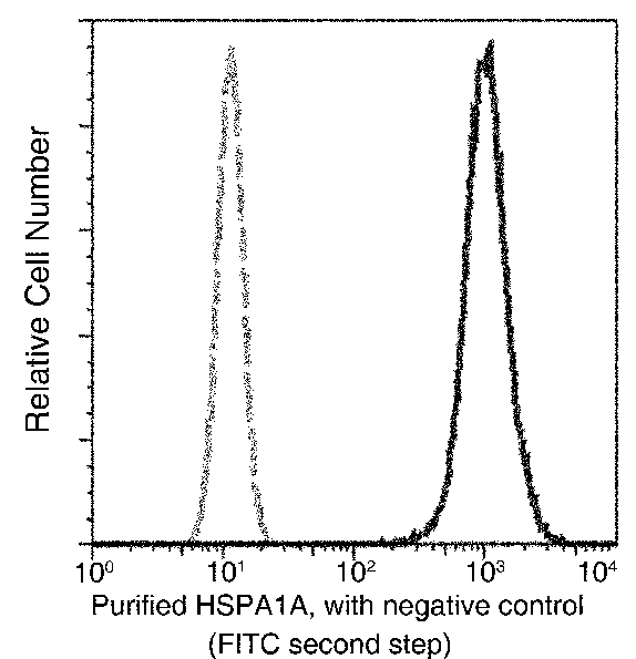
Flow cytometric analysis of Human HSPA1A expression on HeLa cells. The cells were treated and stained with purified anti-Human HSPA1A, then a FITC-conjugated second step antibody. The fluorescence histograms were derived from gated events with the forward and side light-scatter characteristics of intact cells.
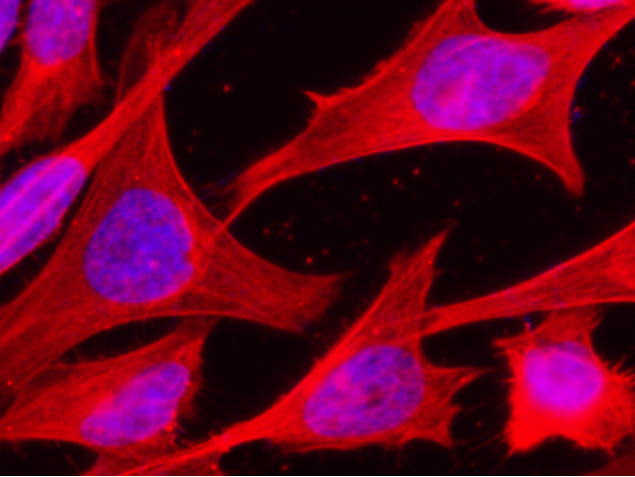
Immunofluorescence staining of Human HSPA1A in Hela cells. Cells were fixed with 4% PFA, permeabilzed with 0.3% Triton X-100 in PBS, blocked with 10% serum, and incubated with rabbit anti-Human HSPA1A monoclonal antibody (1:60) at 37℃ 1 hour. Then cells were stained with the Alexa Fluor® 594-conjugated goat Anti-rabbit IgG secondary antibody (red) and counterstained with DAPI (blue). Positive staining was localized to cytoplasm.
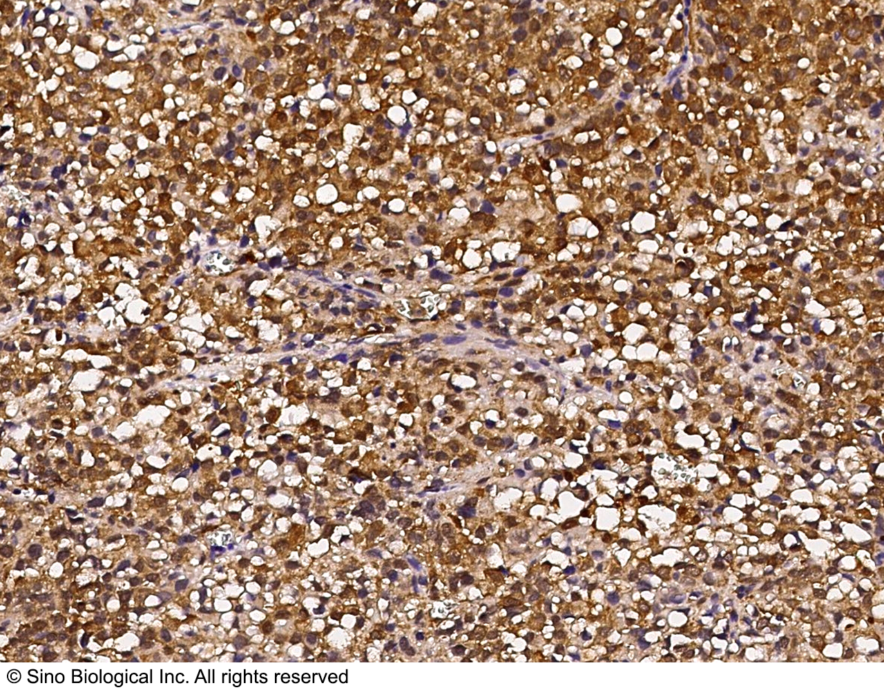
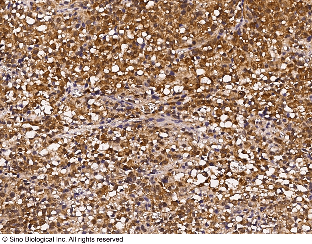
Immunochemical staining of HSPA1A in human hepatoma with rabbit monoclonal antibody (1:1000, formalin-fixed paraffin embedded sections).

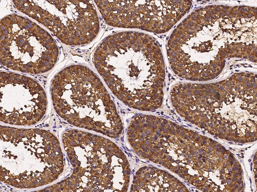
Immunochemical staining of HSPA1A in cynomolgus macaque testis with rabbit monoclonal antibody (1:1000, formalin-fixed paraffin embedded sections).

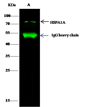
HSPA1A was immunoprecipitated using: Lane A:0.5 mg Hela Whole Cell Lysate, 2 µL anti-HSPA1A rabbit monoclonal antibody and 15 μl of 50 % Protein G agarose. Primary antibody: Anti-HSPA1A rabbit monoclonal antibody,at 1:200 dilution Secondary antibody: Dylight 800-labeled antibody to rabbit IgG (H+L), at 1:5000 dilution. Developed using the odssey technique. Performed under reducing conditions. Observed band size: 70 kDa.
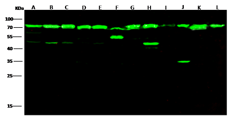
Anti-HSPA1A rabbit monoclonal antibody at 1:500 dilution. Lane A: A549 whole cell lysate. Lane B: HCT116 whole cell lysate. Lane C: Hela whole cell lysate. Lane D: HepG2 whole cell lysate. Lane E: HL60 whole cell lysate. Lane F: A431 whole cell lysate. Lane G: MCF7 whole cell lysate. Lane H: K562 whole cell lysate. Lane I: C6 whole cell lysate. Lane J: 293T whole cell lysate. Lane K: MOLT4 whole cell lysate. Lane L: Jurkat whole cell lysate. Lysates/proteins at 30 μg per lane. Secondary Goat Anti-Rabbit IgG H&L (Dylight800) at 1/10000 dilution. Developed using the Odyssey technique. Performed under reducing conditions. Observed band size:70 kDa.


| Regulatory Status |
RUO – Research Use Only |
|---|
Last modified: September 17, 2024