Widely cited Endoplasmic Reticulum staining with minimal toxicity to cells
- Specifically designed for use with GFP-expressing cell lines, as well as cells expressing blue, cyan or yellow fluorescent proteins (BFPs, CFPs, YFPs)
- Suitable for use with live or post-fixed cells
- Highly resistant to photo-bleaching, concentration quenching and photoconversion
- Suitable for use in conjunction with fluorescein- or coumarin-labeled antibodies
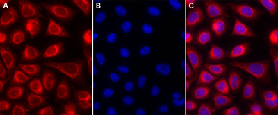
Enzo Life Sciences ER-ID® Red assay kit (GFP-CERTIFIED®) contains a red-emitting, cell-permeable small molecule organic probe to stain Endoplasmic Reticulum (ER) in live cell, or fixed cells. The dye emits in the Texas Red region of the visible light spectrum and can be readily used in combination with other common UV and visible light excitable organic fluorescent dyes/fluorescent proteins in multi-color imaging and detection applications. The kit includes also the Hoechst 33342 dye for the nuclear staining.
Shipping: Available products typically ship within 24/48h, via priority shipping.
Do you need support? Contact Customer Service or Technical Support.
Online Account
Access or Create Your Account
Product Details
Alternative Name |
Endoplasmic reticulum / Organelle |
---|---|
Application |
Fluorescence microscopy |
Application Notes |
Enzo Life Sciences’ ER-ID® Red assay kit (GFP-CERTIFIED®) contains a novel endoplasmic reticulum-selective dye suitable for live cell, or detergent-permeabilized aldehyde-fixed cell staining. |
Contents |
ER-ID® Red detection reagent: 50μl |
Quality Control |
A sample from each lot of ER-ID® Red assay kit is used to stain HeLa cells, using the procedures described in the user manual. The selectivity of the ER-ID® Red dye is evident. |
Quantity |
500 assays |
Technical Info / Product Notes |
The ER-ID® Red assay kit (GFP-CERTIFIED®) is a member of the CELLESTIAL® product line, reagents and assay kits comprising fluorescent molecular probes that have been extensively benchmarked for live cell analysis applications. CELLESTIAL® reagents and kits are optimal for use in demanding cell analysis applications involving confocal microscopy, flow cytometry, microplate readers and HCS/HTS, where consistency and reproducibility are required. |
Handling & Storage
Use/Stability |
With proper storage, the kit components are stable up to the date noted on the product label. Store kit at -20°C in a non-frost free freezer, or -80°C for longer term storage. |
---|---|
Handling |
Protect from light. Avoid freeze/thaw cycles. |
Short Term Storage |
-20°C |
Long Term Storage |
-80°C |
Shipping |
Blue Ice |
Regulatory Status |
RUO – Research Use Only |
---|
- CDNF rescues human iPSCs-derived dopamine neurons through direct binding to unfolded protein response sensors PERK and IRE1α: Kovaleva, V., Shpironok, O., et al.; bioRxiv , (2024)
- MANF regulates neuronal survival and UPR through its ER-located receptor IRE1α: Kovaleva, V., Yu, L. Y., et al.; Cell Rep. 42, 112066 (2023), Abstract
- DIAPH1-MFN2 interaction regulates mitochondria-SR/ER contact and modulates ischemic/hypoxic stress: G. Yepuri, et al.; Nat. Commun. 14, 6900 (2023), Abstract
- CRISPR-Screen Identifies ZIP9 and Dysregulated Zn2+ Homeostasis as a Cause of Cancer-Associated Changes in Glycosylation: T.B. Rømer, et al.; Glycobiology , (2023), Abstract
- Inhibition of TRPA1 Ameliorates Periodontitis by Reducing Periodontal Ligament Cell Oxidative Stress and Apoptosis via PERK/eIF2α/ATF-4/CHOP Signal Pathway: Liu, Q., Guo, S., et al.; Oxid. Med. Cell. Longev. 2022, 4107915 (2022), Abstract
- Secretion of a low‐molecular‐weight species of endogenous GRP94 devoid of the KDEL motif during endoplasmic reticulum stress in Chinese hamster ovary cells: A. Samy, et al.; Traffic 22, 425 (2021), Abstract
- Norcantharidin combined with paclitaxel induces endoplasmic reticulum stress mediated apoptotic effect in prostate cancer cells by targeting SIRT7 expression: M.H. Wu, et al.; Environ. Toxicol. 36, 2206 (2021), Abstract
- MANF regulates unfolded protein response and neuronal survival through its ER-located receptor IRE1α: Kovaleva, V., Yu, L., et al.; bioRxiv , (2020)
- Decoy receptor 3 promotes preosteoclast cell death via reactive oxygen species-induced fas ligand expression and the IL-1α/IL-1 receptor antagonist pathway: Y.J. Peng, et al.; Mediators. Inflamm. 2020, 1237281 (2020), Application(s): Fluorescence microscopy, Abstract — Full Text
- TRPV1 antagonist DWP05195 induces ER stress-dependent apoptosis through the ROS-p38-CHOP pathway in human ovarian cancer cells: Y.Y. Wang, et al.; Cancers (Basel) 12, 1702 (2020), Application(s): Flow cytometry and fluorescence microscopy, Abstract — Full Text
- Effect of 4-phenylbutyrate and valproate on dominant mutations of WFS1 gene in Wolfram syndrome: K. Batjargal, et al.; J. Endocrinol. Invest. 43, 1317 (2020), Abstract
- Differential effects of reactive oxygen species on IgG versus IgM levels in TLR-stimulated B cells: K.M. Gilljam, et al.; J. Immunol. 204, 2133 (2020), Application(s): Flow cytometry, Abstract
- CXCR2 specific endocytosis of immunomodulatory peptide LL-37 in human monocytes and formation of LL-37 positive large vesicles in differentiated: Z. Zhang, et al.; Bone Rep. 12, 100237 (2019), Application(s): Fluorescence microscopy, Abstract — Full Text
- Analysis of intracellular IgG secretion in Chinese hamster ovary cells to improve IgG production: K. Kaneyoshi, et al.; J. Biosci. Bioeng. 127, 107 (2019), Application(s): Fluorescence microscopy, Abstract
- Secretion analysis of intracellular “difficult-to-express” immunoglobulin G (IgG) in Chinese hamster ovary (CHO) cells: K. Kaneyoshi, et al.; Cytotechnology 71, 305 (2019), Application(s): Fluorescence microscopy, Abstract — Full Text
- Variations of Intracellular CaMobilization Initiated by Nanosecond and Microsecond Electrical Pulses in HeLa Cells: N. Ohnishi, et al.; IEEE Trans. Biomed. Eng. 66, 2259 (2019), Abstract
- Inhibition of eIF2α dephosphorylation accelerates pterostilbene-induced cell death in human hepatocellular carcinoma cells in an ER stress and autophagy-dependent manner: C.L. Yu, et al.; Cell Death Dis. 10, 418 (2019), Abstract — Full Text
- Application of Imaging Flow Cytometry for the Characterization of Intracellular Attributes in Chinese Hamster Ovary Cell Lines at the Single-Cell Level: E. Pekle, et al.; Biotechnol. J. 14, e1800675 (2019), Application(s): High-throughput analysis of CHO cells with flow cytometry, Abstract
- A pharmacochaperone-based high-throughput screening assay for the discovery of chemical probes of orphan receptors: C.J. Morfa, et al.; Assay Drug Dev. Technol. 16, 384 (2018), Application(s): Fluorescence microscopy, Abstract — Full Text
- Induction of endoplasmic reticulum stress and mitochondrial dysfunction dependent apoptosis signaling pathway in human renal cancer cells by norcantharidin: M.H. Wu, et al.; Oncotarget 9, 4787 (2018), Application(s): Fluorescence microscopy, Abstract — Full Text
- Protodioscin induces apoptosis through ROS-mediated endoplasmic reticulum stress via the JNK/p38 activation pathways in human cervical cancer cells: C.L. Lin, et al.; Cell. Physiol. Biochem. 46, 322 (2018), Application(s): Flow cytometry and fluorescence microscopy, Abstract
- Domain architecture of vasohibins required for their chaperone-dependent unconventional extracellular release: T. Kadonosono, et al.; Protein Sci. 26, 452 (2017), Abstract
- AT-101 simultaneously triggers apoptosis and a cytoprotective type of autophagy irrespective of expression levels and the subcellular localisation of Bcl-xL and Bcl-2 in MCF7 cells: P. Antonietti, et al.; Biochim. Biophys. Acta 1863, 499 (2016), Application(s): Fluorescence microscopy, Abstract
- Biogenesis of the crystalloid organelle in Plasmodium involves microtubule-dependent vesicle transport and assembly: S. Saeed, et al.; Int. J. Parasitol. 45, 537 (2015), Application(s): Microscopy, Abstract
- Synthesis of a peptide that can translocate to the endoplasmic reticulum: K. Nakatsu, et al.; Biochem. Biophys. Res. Commun. 460, 328 (2015), Application(s): Fluorescent Assay, Abstract
- Targeted protein destabilization reveals an estrogen-mediated ER stress response: K. Raina, et al.; Nat. Chem. Biol. 10, 957 (2014), Application(s): Confocal microscopy, Abstract — Full Text
- Target identification of grape seed extract in colorectal cancer using drug affinity responsive target stability (DARTS) technique: role of endoplasmic reticulum stress response proteins: M.M. Derry, et al.; Curr. Cancer Drug Targets 14, 323 (2014), Abstract
- Molecular cloning and tumour suppressor function analysis of canine REIC/Dkk-3 in mammary gland tumors: K. Ochiai, et al.; Vet. J. 197, 769 (2013), Application(s): ER staining in dog mammary carcinoma cells, Abstract
- Knockdown of RyR3 enhances adiponectin expression through an atf3-dependent pathway: S.H. Tsai, et al.; Endocrinology 154, 1117 (2013), Abstract
- Snowflake vitreoretinal degeneration (SVD) mutation R162W provides new insights into Kir7.1 ion channel structure and function: B.R. Pattnaik, et al.; PLoS One 8, e71744 (2013), Application(s): ER staining in CHO-K1 cells, Abstract — Full Text
- Enhanced optineurin E50K-TBK1 interaction evokes protein insolubility and initiates familial primary open-angle glaucoma: Y. Minegishi, et al.; Hum. Mol. Genet. 22, 3559 (2013), Abstract
Related Products
LYSO-ID® Red detection kit (GFP-CERTIFIED®)
ENZ-51005
Acidic organelle-selective dye for live cell staining of lysosomes

Application | Fluorescence microscopy, Fluorescent detection |
---|
MITO-ID® Green detection kit
ENZ-51022
Photostable, non-toxic and selective mitochondrial dye that stains regardless of membrane potential

Application | Fluorescence microscopy, Fluorescent detection |
---|

Application | Fluorescence microscopy |
---|
GOLGI ID® Green assay kit
ENZ-51028
Dependably localizes to Golgi apparatus with minimal staining of ER

Application | Fluorescence microscopy |
---|
Datasheet, Manuals, SDS & CofA
Certificate of Analysis
Please enter the lot number as featured on the product label
SDS
Enzo Life Science provides GHS Compliant SDS
If your language is not available please fill out the SDS request form