 Lab Essentials
 AMPIVIEW® RNA probes
 Enabling Your Projects
  GMP Services
 Bulk Solutions
 Research Travel Grant
 Have You Published Using an Enzo Product?
A no-transfection, quantitative assay for monitoring autophagy in live cells
- Rapid, no transfection required
- Protocol validated with known inhibitors and activators of autophagic activity
- Selective and comprehensive staining allows differentiation between autophagic flux and autophagolysosome accumulation
- Negligible staining of lysosomes reduces background seen with other dyes
- Facilitates high-throughput screening of activators and inhibitors of autophagy
Enzo Life Sciences CYTO-ID® Autophagy Detection Kit measures autophagic vacuoles and monitors autophagic flux in lysosomally inhibited live cells using a novel dye that selectively labels accumulated autophagic vacuoles. The 488nm-excitable green dye has been optimized through the identification of titratable functional moieties that allow for minimal staining of lysosomes while exhibiting bright fluorescence upon incorporation into pre-autophagosomes, autophagosomes, and autolysosomes (autophagolysosomes). The kit also includes the Hoechst 33342 dye for the nuclear staining, an Autophagy Inducer (Rapamycin) and a Lysosomal Inhibitor (Chloroquine).
Mechanism of Action
The probe is a cationic amphiphilic tracer (CAT) dye that rapidly partitions into cells in a similar manner as drugs that induce phospholipidosis. Careful selection of titratable functional moieties on the dye prevents its accumulation within lysosomes, but enables labeling of vacuoles associated with the autophagy pathway.
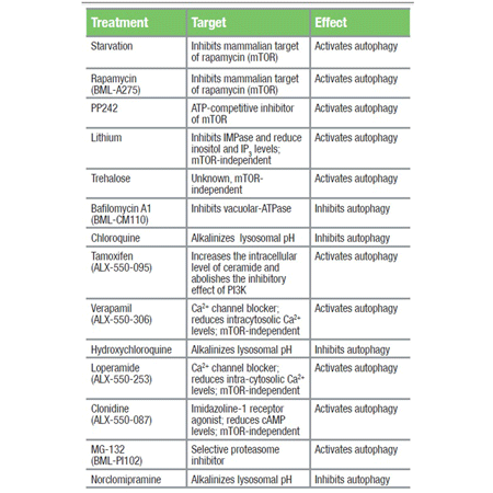
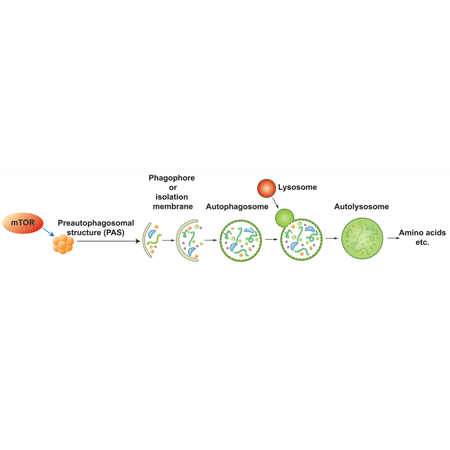
Autophagy is a stress-induced protective mechanism. Less active under basal conditions, the mechanism is utilized by eukaryotic cells through lysosome-mediated bulk degradation of cellular contents when subjected to certain hostile conditions such as nutrient depletion and chemical or environmental stress. The role of increased autophagic activity in the pathology of cancer, neurodegeneration, cardiovascular disease and diabetes has become widely recognized and commonly studied. Induction of autophagic flux can be visualized by enhanced accumulation of autophagic vesicles if lysosomal function is inhibited, preventing removal of these vesicles.
Shipping: Available products typically ship within 24/48h, via priority shipping.
Do you need support? Contact Customer Service or Technical Support.
Online Account
Access or Create Your Account

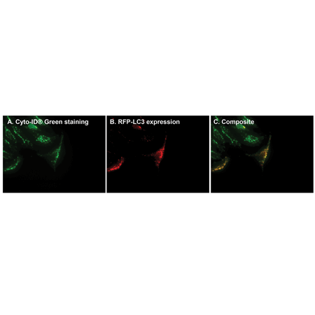
Time-saving, rapid and comprehensive labeling of autophagic vacuoles without transfection. For the purpose of demonstrating advantages of CYTO-ID® Green detection reagent, HeLa cells were first transfected with RFP-LC3 expression vector, treated with 10 µM Tamoxifen overnight, then stained with CYTO-ID® Green detection reagent. Unlike overnight transfection-based assays, the CYTO-ID® Green detection reagent approach labels 100% of cells in 15-30 minutes. Panel A: Green signal indicating CYTO-ID® Green staining of autophagic vesicles; Panel B: RFP-LC3 expression (red) in a subset of successfully transfected cells; Panel C: Composite image, showing CYTO-ID® Green dye-labeled vesicles co-localize with LC3, a specific marker of autophagosomes.

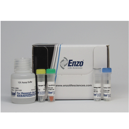
CYTO-ID® Autophagy Detection Kit

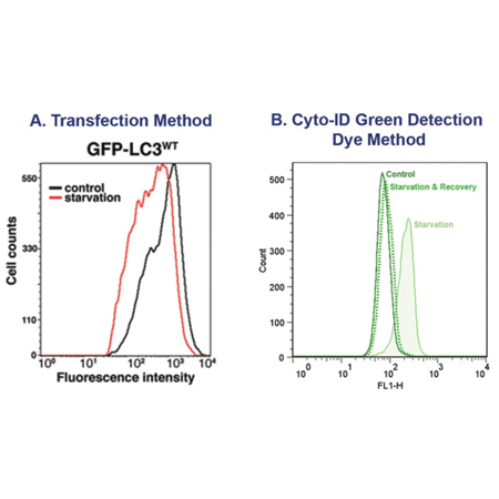
Profile autophagy without transfection. Figure 1A: CHO cells stably expressing GFP-LC3 transfected cell lines results in relatively poor baseline separation of control-vs-starved cell populations, making quantification of autophagy difficult. Figure adapted from Shvets E, Fass E, Elazar Z. Figure 1B: The CYTO-ID® Autophagy Detection Kit specifically labels autophagic vacuoles independent of LC3 protein and eliminates the need for transfection. HeLa cells were subjected to starvation and recovery and then labeled with CYTO-ID® Green detection reagent. The dye enables clear detection and quantification of autophagic and pre-autophagic vacuoles that directly correlates to induction of autophagy.

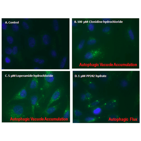
Visualization of autophagic accumulation and autophagic flux. Autophagic vacuole accumulation and flux are both detected by CYTO-ID® Autophagy Green dye as observed by fluorescence microscopy. HeLa cells were mock-induced with 0.2% DMSO (A ) or induced with 100 uM Clonidine hydrochloride (B), 5 uM Loperamide hydrochloride (C ) or 1 uM PP242 hydrate (D) for 12 hours at 37°C. After treatment, cells were incubated with CYTO-ID® Green Detection reagent for 10 min at 37°C and then washed with assay buffer. Nuclei were counter-stained in blue with Hoechst 33342 dye.

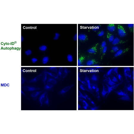
Eliminate background resulting from non-specific lysosomal staining. CYTO-ID® Green dye eliminates background staining of lysosomes seen with other lysosomotrophic dye-based assays that utilize monodansylcadaverine (MDC) (bottom panel). The CYTO-ID® Autophagy kit eliminates the need for a 350 nm UV laser for live cell analysis, and is compatible for use with Hoechst dyes for co-labeling in microscopy applications.

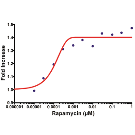
Overnight incubation of HepG2 cells with Rapamycin, an inhibitor of mTOR kinase, results in an increase in CYTO-ID® dye signal.

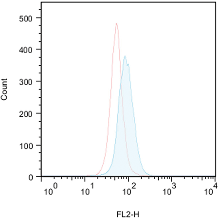
Flow cytometry-based profiling of autophagy with CYTO-ID® Autophagy Detection Kit: Control (red-lined peak) uninduced and 10uM Tamoxifen (ALX-550-095) treated (blue-filled peak) Jurkat cells (T-cell leukemia) were used. After 18 hours treatment, cells were loaded with CYTO-ID® Green Detection Reagent, then analyzed without washing by flow cytometry. Results are presented by histogram overlays. Control cells were stained as well but mostly display low fluorescence. In the samples treated with 10uM Tamoxifen for 18 hours, CYTO-ID® Green dye signal increases about 2-fold, indicating that Tamoxifen causes an increase in autophagy in Jurkat cells.

Flow cytometry-based profiling of autophagy with CYTO-ID® Autophagy Detection Kit: Control (red-lined peak) uninduced and 10uM Tamoxifen (ALX-550-095) treated (blue-filled peak) Jurkat cells (T-cell leukemia) were used. After 18 hours treatment, cells were loaded with CYTO-ID® Green Detection Reagent, then analyzed without washing by flow cytometry. Results are presented by histogram overlays. Control cells were stained as well but mostly display low fluorescence. In the samples treated with 10uM Tamoxifen for 18 hours, CYTO-ID® Green dye signal increases about 2-fold, indicating that Tamoxifen causes an increase in autophagy in Jurkat cells.

Schematic depiction of autophagy. Cytosolic material is sequestered by an expanding membrane sac, the phagophore, resulting in the formation of a double-membrane vesicle, an autophagosome. The outer membrane of the autophagosome subsequently fuses with the lysosome, and the internal material is degraded in the autolysosome. Various regulators of autophagy are also depicted in the diagram.
Product Details
| Application |
Flow Cytometry, Fluorescence microscopy, Fluorescent detection, HTS |
|---|---|
| Application Notes |
The CYTO-ID® Autophagy detection kit provides a rapid, specific and quantitative approach for monitoring autophagy in live cells by fluorescence microscopy, flow cytometry, and microplate reader. |
| Contents |
CYTO-ID® Green Detection Reagent |
| Quality Control |
A sample from each lot of CYTO-ID® Autophagy detection kit is used to stain HeLa Cells as described in user manual. CYTO-ID® autophagy detection reagent is incorporated into induced cells, observed as accumulative typical spherical vacuoles in foci or throughout cytoplasm. Comparing to untreated HeLa cells, treated sample demonstrate significant increase in fluorescence under microscope. |
| Quantity |
For -K200 size: For -0050 size: |
| Technical Info / Product Notes |
The CYTO-ID® Autophagy Detection kit is a member of the CELLESTIAL® product line, reagents and assay kits comprising fluorescent molecular probes that have been extensively benchmarked for live cell analysis applications. Featured in: Application Notes: A Novel Image-Based Cytometry Method for Autophagy Detection in Living Cells Visualizing subcellular vesicles to quantitate autophagy in neuronal cells Cited samples: |
Handling & Storage
| Use/Stability |
With proper storage, the kit components are stable for one year from date of receipt. |
|---|---|
| Handling |
Protect from light. Avoid freeze/thaw cycles. |
| Short Term Storage |
-20°C |
| Long Term Storage |
-80°C |
| Shipping |
Blue Ice |
| Regulatory Status |
RUO – Research Use Only |
|---|
- Neuroglobin regulates autophagy through mTORC1/RAPTOR/ULK-1 pathway in human neuroblastoma cells: Manganelli, V., Costanzo, M., et al.; Sci. Rep. 15, 7642 (2025), Abstract
- A sesquiterpene lactone, tomentosin, as a novel anticancer agent: orchestrating apoptosis, autophagy, and ER stress in colorectal cancer: S. Çetinkaya, et al.; Naunyn Schmiedebergs Arch Pharmacol , (2025), Abstract
- Ancient genomic linkage of α-globin and Nprl3 couples metabolism with erythropoiesis: A.E. Preston, et al.; Nat Commun 16, 2749 (2025), Abstract
- Bioengineering the metabolic network of CAR T cells with GLP-1 and Urolithin A increases persistence and long-term anti-tumor activity: A. Akhtar, et al.; Cell Rep Med 6, 102021 (2025), Abstract
- Hypoxia‑induced SREBP1‑mediated lipogenesis and autophagy promote cell survival via fatty acid oxidation in breast cancer cells: J.H. Jung, et al.; Oncol Lett 29, 175 (2025), Abstract
- Immunomodulatory Nanoparticles Induce Autophagy in Macrophages and Reduce Mycobacterium tuberculosis Burden in the Lungs of Mice: R.B. Bekale, et al.; ACS Infect Dis 11, 610 (2025), Abstract
- Mitochondrial Mayhem: How cigarette smoke induces placental dysfunction through MMS19 degradation: P. Zhou, et al.; Ecotoxicol Environ Saf 290, 117728 (2025), Abstract
- Sauchinone Ameliorates Senescence Through Reducing Mitochondrial ROS Production: M.U. Kuk, et al.; Antioxidants 14, 259 (2025), Abstract
- Waterborne ammonia toxicity damages crustacean hemocytes via lysosome-dependent autophagy: A case study of swimming crabs Portunus trituberculatus: Y. Lu, et al.; Environ Res 272, 120985 (2025), Abstract
- Enhanced cytotoxicity of T-DM1 in HER2-low carcinomas via autophagy inhibition: Zhang, J., Chang, X., et al.; PLoS One 20, e0322029 (2025), Abstract
- Imbalanced TGFβ signalling and autophagy drive erythroid priming of hematopoietic stem cells in β-thalassemia: Lidonnici, M. R., Chianella, G., et al.; Nat. Commun. 16, 5639 (2025), Abstract
- Galectin-9 treatment is cytotoxic for B cell lymphoma by disrupting autophagy: Koll, L., Lourens, H. J., et al.; Front. Pharmacol. 16, 1601235 (2025), Reactant(s): Human, Abstract
- Inhibiting CD36 palmitoylation improves cardiac function post-infarction by regulating lipid metabolic homeostasis and autophagy: Zhang, Q., Li, J., et al.; Nat. Commun. 16, 6602 (2025), Abstract
- Distinctive Gene Expression Profiles and Biological Responses of Skin Fibroblasts to Nicotinamide Mononucleotide: Implications for Longevity Effects on Skin.: Kang, S., Park, J., et al.; Biomedicines 13, 2395 (2025), Abstract
- Unc-51 Like Kinase 3 (ULK3) is essential for autophagy and cell survival in multiple myeloma: Lynch, C., Tauro, M., et al.; Research Square , (2025)
- IGF2BP1/IMP1 Deletion Enhances a Facultative Stem Cell State via Regulation of MAP1LC3B: L.R. Parham, et al.; Cell. Mol. Gastroenterol. Hepatol. 17, 439 (2024), Abstract
- JS-K activates G2/M checkpoints through the DNA damage response and induces autophagy via CAMKKβ/AMPKα/mTOR pathway in bladder cancer cells: Y. Zhao, et al.; J. Cancer 15, 343 (2024), Abstract
- RhoB expression associated with chemotherapy response and prognosis in colorectal cancer: M. Kopsida, et al.; Cancer Cell Int. 24, s12935 (2024), Abstract
- Indocyanine green based photodynamic therapy for keloids: Fundamental investigation and clinical improvement: J. Shao, et al.; Photodiagnosis Photodyn. Ther. 45, 103903 (2024), Abstract
- µMap proximity labeling in living cells reveals stress granule disassembly mechanisms: C.R. Pan, et al.; Nat. Chem. Biol. , (2024), Abstract
- Autophagy Contributes to Homeostasis in Esophageal Epithelium Where High Autophagic Vesicle Level Marks Basal Cells With Limited Proliferation and Enhanced Self-Renewal Potential: Klochkova, A., Karami, A. L., et al.; Cell. Mol. Gastroenterol. Hepatol. 18, 15 (2024), Abstract
- Cisplatin and Starvation Differently Sensitize Autophagy in Renal Carcinoma: A Potential Therapeutic Pathway to Target Variegated Drugs Resistant Cancerous Cells: Dutta, A., Thakur, S., et al.; Cells 13, (2024), Abstract
- Enhancing antitumor efficacy of CLDN18.2-directed antibody-drug conjugates through autophagy inhibition in gastric cancer: Xue, W., Xu, C., et al.; Cell Death Discov. 10, 393 (2024), Abstract
- Identification of Cellular Isoschaftoside-Mediated Anti-Senescence Mechanism in RAC2 and LINC00294: Lee, Y. H., So, B. H., et al.; Molecules 29, (2024), Abstract
- Induction of Multiple Alternative Mitogenic Signaling Pathways Accompanies the Emergence of Drug-Tolerant Cancer Cells: Celeste, F. V., Powers, S., et al.; Cancers (Basel) 16, (2024), Abstract
- Lamin B1 curtails early human papillomavirus infection by safeguarding nuclear compartmentalization and autophagic capacity: Molenberghs, F., Verschuuren, M., et al.; Cell. Mol. Life Sci. 81, 141 (2024), Abstract
- Reduced anti-Müllerian hormone action in cumulus-oocyte complexes is beneficial for oocyte maturation without affecting oocyte competence: Xu, F., Bagnjuk, K., et al.; Front. Endocrinol. (Lausanne) 15, 1365260 (2024), Abstract
- Senescence Rejuvenation through Reduction in Mitochondrial Reactive Oxygen Species Generation by Polygonum cuspidatum Extract: In Vitro Evidence: Yoon, J. H., Kim, Y. H., et al.; Antioxidants (Basel) 13, (2024), Abstract
- 5-Fluorouracil induces apoptosis in nutritional deprived hepatocellular carcinoma through mitochondrial damage: A. Dutta, et al.; Sci Rep 14, 23387 (2024), Abstract
- Cysteine Leukotriene Receptor Antagonist-Montelukast Effects on Diabetic Retinal Microvascular Endothelial Cells Curtail Autophagy: A.M. Awad, et al.; Invest Ophthalmol Vis Sci 65, 15 (2024), Abstract
- Neuroprotective Assessment of Nutraceutical (Betanin) in Neuroblastoma Cell Line SHSY-5Y: An in-Vitro and in-Silico Approach: K. Natarajan, et al.; Neurochem Res 50, 54 (2024), Abstract
- The Survival of Human Intervertebral Disc Nucleus Pulposus Cells under Oxidative Stress Relies on the Autophagy Triggered by Delphinidin: M.E. Bahar, et al.; Antioxidants 13, 759 (2024), Abstract
- Deciphering MARCH5’s impact on multiple myeloma: insights into autophagy regulation and AKT-FOXO3 signaling: Bashiri, H., Khalilnezhad, A., et al.; Blood Neoplasia 1, 100046 (2024), Abstract
- Old age alters inflammation and autophagy signaling in the brain, leading to exacerbated neurological outcomes after spinal cord injury in male mice: Lei, Z., Ritzel, R. M., et al.; Brain Behav. Immun. 120, 439 (2024), Abstract
- Modulating autophagy to boost the antitumor efficacy of TROP2-directed antibody-drug conjugate in pancreatic cancer.: Xu, C., Huang, X., et al.; Biomed. Pharmacother. 180, 117550 (2024), Abstract
- Targeting Annexin A1 as a Druggable Player to Enhance the Anti-Tumor Role of Honokiol in Colon Cancer through Autophagic Pathway: X. Wang, et al.; Pharmaceuticals 16, 70 (2023), Abstract
- Amorfrutin B Protects Mouse Brain Neurons from Hypoxia/Ischemia by Inhibiting Apoptosis and Autophagy Processes Through Gene Methylation- and miRNA-Dependent Regulation: K. Przepiórska, et al.; Mol. Neurobiol. 60, 576 (2023), Abstract
- Brain injury accelerates the onset of a reversible agerelated microglial phenotype associated with inflammatory neurodegeneration: R.M. Ritzel, et al.; Sci. Adv. 9, eadd1101 (2023), Abstract
- Functional restoration of lysosomes and mitochondria through modulation of AKT activity ameliorates senescence: M.U. Kuk, et al.; Exp. Gerontol. 173, 112091 (2023), Abstract
- Impaired Autophagy in Krabbe Disease: The Role of BCL2 and Beclin-1 Phosphorylation: N. Papini, et al.; Int. J. Mol. Sci. 24, 5984 (2023), Abstract
- Nanoparticles of folic acid-methyl-b-cyclodextrin (FA-MbCD)/adamantane-albumin exhibit enhanced antitumor activity compared with FA-MbCD alone: A. Sakai, et al.; FEBS Open Bio 13, 233 (2023), Abstract
- Novel hydroxamic acid derivative induces apoptosis and constrains autophagy in leukemic cells: M.A. Fischer, et al.; J. Adv. Res. , (2023), Abstract
- Surface-Modified Inhaled Microparticle-Encapsulated Celastrol for Enhanced Efficacy in Malignant Pleural Mesothelioma: X. Wang, et al.; Int. J. Mol. Sci. 24, 5204 (2023), Abstract
- Syk regulates the haemocyte autophagy through inducing the mRNA expressions of autophagy-related genes and the cleavage of CgLC3 in oyster antibacterial immunity: H. Yao, et al.; Fish Shellfish Immunol. Rep. 4, 100085 (2023), Abstract
- BR101801 enhances the radiosensitivity of p53-deficient colorectal cancer cells by inducing G2/M arrest, apoptosis, and senescence in a p53-independent manner: M. Park, et al.; Am. J. Cancer Res. 13, 5887 (2023), Abstract
- Low extracellular magnesium induces phenotypic and metabolic alterations in C2C12-derived myotubes: M. Zocchi, et al.; Sci. Rep. 13, 19425 (2023), Abstract
- Development of a bioengineered Erwinia chrysanthemi asparaginase to enhance its anti-solid tumor potential for treating gastric cancer: S.Y. Tim, et al.; Int. J. Biol. Macromol. 253, 127742 (2023), Abstract
- Isoquercitrin from Apocynum venetum L. Exerts Antiaging Effects on Yeasts via Stress Resistance Improvement and Mitophagy Induction through the Sch9/Rim15/Msn Signaling Pathway: Y. Liu, et al.; Antioxidants 12, 1939 (2023), Abstract
- A novel combination therapy with Cabozantinib and Honokiol effectively inhibits c-Met-Nrf2-induced renal tumor growth through increased oxidative stress: L. Rawat, et al.; Redox Biol. 68, 102945 (2023), Abstract
- Adipose Mesenchymal Stem Cell-Derived Exosomes Promote the Regeneration of Corneal Endothelium Through Ameliorating Senescence: Y. Ryu, et al.; Invest. Ophthalmol. Vis. Sci. 64, 29 (2023), Abstract
- Restoration of PM2.5-induced spermatogonia GC-1 cellular damage by parthenolide via suppression of autophagy and inflammation: An in vitro study: H.J. Gu, et al.; Toxicology 499, 153651 (2023), Abstract
- Age-related changes in plasma extracellular vesicles influence neuroinflammation in the brain and neurological outcome after traumatic spinal cord injury: Lei, Z., Krishnamachary, B., et al.; Research Square , (2023)
- Autolysosomal activation combined with lysosomal destabilization efficiently targets myeloid leukemia cells for cell death: Shah, H., Stankov, M., et al.; Front. Oncol. 13, 999738 (2023), Abstract
- Autophagy contributes to homeostasis in esophageal epithelium where high autophagic vesicle content marks basal cells with limited proliferation and enhanced self-renewal potential: Klochkova, A., Karami, A. L., et al.; bioRxiv , (2023)
- Development of small fluorescent probes for the analysis of autophagy kinetics: Sakurai, H. T., Iwashita, H., et al.; iScience 26, 107218 (2023), Abstract
- Dynamin-2 deficiency causes age- and sex-dependent neutropenia and myelodysplasia in mice: Willis, A. J., Corey, S. J., et al.; Blood Adv. 7, 1418 (2023), Abstract
- Fasting-Mimicking Diet Inhibits Autophagy and Synergizes with Chemotherapy to Promote T-Cell-Dependent Leukemia-Free Survival: Buono, R., Tucci, J., et al.; Cancers (Basel) 15, (2023), Abstract
- Fisetin induces apoptosis in colorectal cancer cells by suppressing autophagy and down-regulating nuclear factor erythroid 2-related factor 2 (Nrf2): Pandey, A., Trigun, S. K., et al.; J. Cell. Biochem. 124, 1289 (2023), Abstract
- Galectin-9 has non-apoptotic cytotoxic activity toward acute myeloid leukemia independent of cytarabine resistance: Choukrani, G., Visser, N., et al.; Cell Death Discov. 9, 228 (2023), Abstract
- Galectin-9 has non-apoptotic cytotoxic activity towards Acute Myeloid Leukemia independent of cytarabine resistance: Choukrani, G., Visser, N., et al.; bioRxiv , (2023)
- Inhibition of autophagy in microglia and macrophages exacerbates innate immune responses and worsens brain injury outcomes: Hegdekar, N., Sarkar, C., et al.; Autophagy 19, 2026 (2023), Abstract
- LAMP2 regulates autophagy in the thymic epithelium and thymic stroma-dependent CD4 T cell development: Rodrigues, P. M., Sousa, L. G., et al.; Autophagy 19, 426 (2023), Abstract
- Lipofuscin Granule Accumulation Requires Autophagy Activation: Song, S. B., Shim, W., et al.; Mol. Cells 46, 486 (2023), Abstract
- Role of a Novel Heparanase Inhibitor on the Balance between Apoptosis and Autophagy in U87 Human Glioblastoma Cells: Manganelli, V., Misasi, R., et al.; Cells 12, (2023), Abstract
- Discovery of a novel dual functional phenylpyrazole-styryl hybrid that induces apoptotic and autophagic cell death in bladder cancer cells: S.W. Leong, et al.; Eur. J. Med. Chem. 115335, 254 (2023), Abstract
- Knockdown of the Autophagy Protein Beclin-1 Does Not Affect Innate Cytokine Production in Human Lung Epithelial Cells during Respiratory Syncytial Virus Infection: K. Parameswaran, et al.; Trop. Med. Infect. Dis. 8, 434 (2023), Abstract
- Distinct roles of core autophagy-related genes in zebrafish definitive hematopoiesis: X.-K. Chen, et al.; Autophagy 20, 830 (2023), Application(s): Staining of whole zebrafish embryos and FACS-sorted zebrafish embryo cells, Abstract — Full Text
- Identification of distinct slow mode of reversible adaptation of pancreatic ductal adenocarcinoma to the prolonged acidic pH microenvironment: T.C. Wu, et al.; J. Exp. Clin. Cancer Res. 41, 137 (2022), Abstract
- Spautin-1 inhibits mitochondrial complex I and leads to suppression of the unfolded protein response and cell survival during glucose starvation: K. Kunimasa, et al.; Sci. Rep. 12, 11533 (2022), Abstract
- GSTO1 confers drug resistance in HCT‑116 colon cancer cells through an interaction with TNFαIP3/A20: S. Paul, et al.; Int. J. Oncol. 61, 137 (2022), Abstract
- Snake venom induces an autophagic cell death via activation of the JNK pathway in colorectal cancer cells: J.E. Yu, et al.; J. Cancer 13, 3333 (2022), Abstract
- BCL2 inhibitor ABT-199 and BCL2L1 inhibitor WEHI-539 coordinately promote NOXA-mediated degradation of MCL1 in human leukemia cells: J.T. Chiou, et al.; Chem. Biol. Interact. 361, 109978 (2022), Abstract
- NEDD4L binds the proteasome and promotes autophagy and bortezomib sensitivity in multiple myeloma: X. Huang, et al.; Cell Death Dis. 13, 197 (2022), Abstract
- Cellular Effects of Cyclodextrins: Studies on HeLa Cells: Á. Rusznyák, et al.; Molecules 27, 1589 (2022), Abstract
- CNS serotonin content mediating food deprivation-enhanced learning is regulated by hemolymph tryptophan concentration and autophagic flux in the pond snail: Y. Totani, et al.; Nutr. Neurosci. , (2022), Abstract
- Regulatory Effects of Astragaloside IV on Hyperglycemia-Induced Mitophagy in Schwann Cells: X. Wei, et al.; Evid. Based Complement. Alternat. Med. 2022, 7864308 (2022), Abstract
- HIF-1α/FOXO1 axis regulated autophagy is protective for β cell survival under hypoxia in human islets: R. Liang, et al.; Biochim. Biophys. Acta Mol. Basis Dis. 1868, 166356 (2022), Abstract
- Magnesium Homeostasis in Myogenic Differentiation – A Focus on the Regulation of TRPM7, MagT1 and SLC41A1 Transporters: M. Zocchi, et al.; Int. J. Mol. Sci. 23, 1658 (2022), Abstract
- Air Plasma-Activated Medium Evokes a Death-Associated Perinuclear Mitochondrial Clustering: M. Suzuki-Karasaki, et al.; Int. J. Mol. Sci. 23, 1124 (2022), Abstract
- Integrative analysis of the miRNA-mRNA regulation network in hemocytes of Penaeus vannamei following Vibrio alginolyticus infection: F. Wang, et al.; Dev. Comp. Immunol. 131, 104390 (2022), Abstract
- A peptide interfering with the dimerization of oncogenic KITENIN protein and its stability suppresses colorectal tumour progression: S.J. Kim et al.; Clin. Transl. Med. 12, e871 (2022), Abstract
- PHF20 is crucial for epigenetic control of starvation-induced autophagy through enhancer activation: S.W. Park, et al.; Nucleic Acids Res. 50, 7856 (2022), Abstract
- miR-23b-3p Modulating Cytoprotective Autophagy and Glutamine Addiction in Sorafenib Resistant HepG2, a Hepatocellular Carcinoma Cell Line: R. Kaur, et al.; Genes 13, 1375 (2022), Abstract
- Activated amino acid response pathway generates apatinib resistance by reprograming glutamine metabolism in non-small-cell lung cancer: X. Zhou, et al.; Cell Death Dis. 13, 636 (2022), Abstract
- Siah-1-interacting protein regulates mutated huntingtin protein aggregation in Huntington’s disease models: E. Latoszek, et al.; Cell Biosci. 12, 34 (2022), Abstract
- Celastrol upregulated ATG7 triggers autophagy via targeting Nur77 in colorectal cancer: W. Zhang, et al.; Phytomedicine 104, 154280 (2022), Abstract
- Autophagy induced by taurolidine protects against polymicrobial sepsis by promoting both host resistance and disease tolerance: J. Huang, et al.; PNAS 119, e2121244119 (2022), Abstract
- Nanoparticle formulation of the fusion protein virus like particles of respiratory syncytial virus stimulates enhanced in vitro antigen presentation and autophagy: I. Menon, et al.; Int. J. Pharm. 623, 121919 (2022), Abstract
- The preventive effect of loganin on oxidative stress-induced cellular damage in human keratinocyte HaCaT cells: C. Park, et al.; Biosci. Trends 16, 291 (2022), Abstract
- Protective autophagy decreases lorlatinib cytotoxicity through Foxo3a-dependent inhibition of apoptosis in NSCLC: C. Lu, et al.; Cell Death Discov. 8, 221 (2022), Abstract
- 3,4,5-O-tricaffeoylquinic acid with anti-radiation activity suppresses LPS-induced NLRP3 inflammasome activation via autophagy in THP-1 macrophages: J. Liu, et al.; Mol. Immunol. 147, 187 (2022), Abstract
- Alcohol Metabolism Enriches Squamous Cell Carcinoma Cancer Stem Cells That Survive Oxidative Stress via Autophagy: M. Shimonosono, et al.; Biomolecules 11, 1479 (2022), Abstract
- Asiatic acid from Cyclocarya paliurus regulates the autophagy-lysosome system via directly inhibiting TGF-β type I receptor and ameliorates diabetic nephropathy fibrosis: X.X. Zhang, et al.; Food Funct. 13, 5536 (2022), Abstract
- Inhibition of B-cell lymphoma 2 family proteins alters optical redox ratio, mitochondrial polarization, and cell energetics independent of cell state: A.A. Gillette, et al.; J. Biomed. Opt. 27, 56505 (2022), Abstract
- Combined Levo-tetrahydropalmatine and diphenyleneiodonium chloride enhances antitumor activity in hepatocellular carcinoma: X. Yin, et al.; Pharmacol. Res. 179, 106219 (2022), Abstract
- Gallium(III) Complex with Cloxyquin Ligands Induces Ferroptosis in Cancer Cells and Is a Potent Agent against Both Differentiated and Tumorigenic Cancer Stem Rhabdomyosarcoma Cells: M. Hreusova, et al.; Bioinorg. Chem. Appl. 2022, 3095749 (2022), Abstract
- Nigrosporins B, a Potential Anti-Cervical Cancer Agent, Induces Apoptosis and Protective Autophagy in Human Cervical Cancer Ca Ski Cells Mediated by PI3K/AKT/mTOR Signaling Pathway: J. Zhang, et al.; Molecules 27, 2431 (2022), Abstract
- Anti-neoplastic sulfonamides alter the metabolic homeostasis and disrupt the suppressor activity of regulatory T cells: R. Gedaly, et al.; Sci. Rep. 12, 19112 (2022), Abstract
- Bee Venom Triggers Autophagy-Induced Apoptosis in Human Lung Cancer Cells via the mTOR Signaling Pathway: J.E. Yu, et al.; J. Oncol. 2022, 8916464 (2022), Abstract
- Bioreactor expansion reconfigures metabolism and extracellular vesicle biogenesis of human adipose-derived stem cells in vitro: R. Jeske, et al.; Biochem Eng. J. 188, 108711 (2022), Abstract
- Camboginol and Morelloflavone from Garcinia dulcis (Roxb.) Kurz Flower Extract Promote Autophagic Cell Death against Human Glioblastoma Cells through Endoplasmic Reticulum Stress: T. Siangcham, et al.; Prev. Nutr. Food Sci. 27, 376 (2022), Abstract
- MiR-302a Regenerates Human Corneal Endothelial Cells against IFN-γ-Induced Cell Death: S.H. Park, et al.; Cells 12, 36 (2022), Abstract
- Oleic acid from cancer-associated fibroblast promotes cancer cell stemness by stearoyl-CoA desaturase under glucose-deficient condition: S.H. Hwang, et al.; Cancer Cell Int. 22, 404 (2022), Abstract
- Phloroglucinol Attenuates DNA Damage and Apoptosis Induced by Oxidative Stress in Human Retinal Pigment Epithelium ARPE-19 Cells by Blocking the Production of Mitochondrial ROS: C. Park, et al.; Antioxidants 11, 2353 (2022), Abstract
- Sexual identity of enterocytes regulates autophagy to determine intestinal health, lifespan and responses to rapamycin: J.C. Regan, et al.; Nat. Aging 2, 1145 (2022), Abstract
- Stevia and Stevioside Attenuate Liver Steatosis through PPARα-Mediated Lipophagy in db/db Mice Hepatocytes: M. Park, et al.; Antioxidants 11, 2496 (2022), Abstract
- Activation of Tumor-Cell STING Primes NK-Cell Therapy: Knelson, E. H., Ivanova, E. V., et al.; Cancer Immunol. Res. 10, 947 (2022), Abstract
- Autophagy-cell death balance is maintained by Polycomb-mediated regulation during stem cell differentiation: Puri, D., Kelkar, A., et al.; bioRxiv , (2022)
- Carbon Monoxide Activates PERK-Regulated Autophagy to Induce Immunometabolic Reprogramming and Boost Antitumor T-cell Function: Chakraborty, P., Parikh, R. Y., et al.; Cancer Res. 82, 1969 (2022), Abstract
- Combined treatment with ruxolitinib and MK-2206 inhibits the JAK2/STAT5 and PI3K/AKT pathways via apoptosis in MDA-MB-231 cells: Celik, E. G., Eroğlu, O., et al.; Research Square , (2022)
- Functional and transcriptional profiling of microglial activation during the chronic phase of TBI identifies an age-related driver of poor outcome in old mice: Ritzel, R. M., Li, Y., et al.; Geroscience 44, 1407 (2022), Abstract
- Impairment of autophagy after spinal cord injury potentiates neuroinflammation and motor function deficit in mice: Li, Y., Lei, Z., et al.; Theranostics 12, 5364 (2022), Abstract
- Induction of Multiple Alternative Mitogenic Signaling Pathways Accompanies Emergence of Slowly Growing Drug-Tolerant Cancer Cells: Celeste, F., Powers, S., et al.; Research Square , (2022)
- Iron regulatory protein (IRP)-mediated iron homeostasis is critical for neutrophil development and differentiation in the bone marrow: Bonadonna, M., Altamura, S., et al.; Sci. Adv. 8, eabq4469 (2022), Abstract
- Metronomic doses and drug schematic combination response tested within chambered coverslips for the treatment of breast cancer cells (JIMT-1): Rosero, G., Pattarone, G., et al.; PLoS One 17, e0274911 (2022), Abstract
- Monitoring Autophagy at Cellular and Molecular Level in Crassostrea gigas During an Experimental Ostreid Herpesvirus 1 (OsHV-1) Infection: Picot, S., Faury, N., et al.; Front. Cell. Infect. Microbiol. 12, 858311 (2022), Abstract
- Selaginella tamariscina Inhibits Glutamate-Induced Autophagic Cell Death by Activating the PI3K/AKT/mTOR Signaling Pathways: Jeong, Y. H., Kim, T. I., et al.; Int. J. Mol. Sci. 23, (2022), Abstract
- Spatially resolved phosphoproteomics reveals fibroblast growth factor receptor recycling-driven regulation of autophagy and survival: Watson, J., Ferguson, H. R., et al.; Nat. Commun. 13, 6589 (2022), Abstract
- Targeting regulation of ATP synthase 5 alpha/beta dimerization alleviates senescence: Lee, Y. H., Choi, D., et al.; Aging (Albany NY) 14, 678 (2022), Abstract
- The role of FYCO1-dependent autophagy in lens fiber cell differentiation: Khan, S. Y., Ali, M., et al.; Autophagy 18, 2198 (2022), Abstract
- β2-adrenergic receptor signaling regulates metabolic pathways critical to myeloid-derived suppressor cell function within the TME: H. Mohammadpour, et al.; Cell Rep. 37, 109883 (2021), Abstract
- Cellular Toxicity Mechanisms and the Role of Autophagy in Pt(IV) Prodrug-Loaded Ultrasmall Iron Oxide Nanoparticles Used for Enhanced Drug Delivery: L.G. Romero, et al.; Pharmaceutics 13, 1730 (2021), Abstract
- Tumor-treating fields as a proton beam-sensitizer for glioblastoma therapy: W.S. Lee, et al.; Am. J. Cancer Res. 11, 4582 (2021), Abstract
- Serum supplementation during bovine embryo culture affects their development and proliferation through macroautophagy and endoplasmic reticulum stress regulation: E.J. Soto-Moreno, et al.; PLoS One 16, e0260123 (2021), Abstract
- Carnosine suppresses human colorectal cancer cell proliferation by inducing necroptosis and autophagy and reducing angiogenesis: S.L. Hsieh, et al.; Oncol. Lett. 23, 44 (2021), Abstract
- Autophagy Promotes Hepatic Cystogenesis in Polycystic Liver Disease via Depletion of Cholangiocyte Ciliogenic Proteins: A.I. Masyuk, et al.; Hepatology , (2021), Abstract
- A Combination therapy using an mTOR inhibitor and Honokiol effectively induces autophagy through the modulation of AXL and Rubicon in renal cancer cells and restricts renal tumor growth following organ transplantation: A. Sabarwal, et al.; Carcinogenesis , (2021), Abstract
- AMPK-Mediated Metabolic Switching Is High Effective for Phytochemical Levo-Tetrahydropalmatine (l-THP) to Reduce Hepatocellular Carcinoma Tumor Growth: X. Yin, et al.; Metabolites 11, 811 (2021), Abstract
- Hematopoiesis under telomere attrition at the single-cell resolution: N. Thongon, et al.; Nat. Commun. 12, 6850 (2021), Abstract
- REV1 Inhibition Enhances Radioresistance and Autophagy: K.E. Ikeh, et al.; Cancers 13, 5290 (2021), Abstract
- Cynaroside protects the blue light-induced retinal degeneration through alleviating apoptosis and inducing autophagy in vitro and in vivo: J.H. Feng, et al.; Phytomedicine 88, 153604 (2021), Abstract
- Leber’s Hereditary Optic Neuropathy Arising From the Synergy Between ND1 3635G>A Mutation and Mitochondrial YARS2 Mutations: X. Jin, et al.; Invest. Ophthalmol. Vis. Sci. 62, 22 (2021), Abstract
- Features of the Population of Mouse Peritoneal Macrophages Isolated after Stimulation with Concanavalin A and Thioglycolate: E.S. Zubkova, et al.; Bull. Exp. Biol. Med. 171, 532 (2021), Abstract
- Alpha1-antitrypsin counteracts heme-induced endothelial cell inflammatory activation, autophagy dysfunction and death: K. Madyaningrana, et al.; Redox Biol. 46, 102060 (2021), Abstract
- Aggregated Tau-PHF6 (VQIVYK) Potentiates NLRP3 Inflammasome Expression and Autophagy in Human Microglial Cells: C. Panda, et al.; Cells 10, 1652 (2021), Abstract
- Advanced Maternal Age Deteriorates the Developmental Competence of Vitrified Oocytes in Mice: J. H. Lee, et al.; Cells 10, 1563 (2021), Abstract
- PLAC8 promotes adriamycin resistance via blocking autophagy in breast cancer: Y. Chen, et al.; J. Cell. Mol. Med. , (2021), Abstract
- Inhibiting autophagy targets human leukemic stem cells and hypoxic AML blasts by disrupting mitochondrial homeostasis: K.M. Dykstra, et al.; Blood Adv. 5, 2087 (2021), Abstract
- Atractylodin inhibited the migration and induced autophagy in cholangiocarcinoma cells via PI3K/AKT/mTOR and p38MAPK signalling pathways: B. Acharya, et al.; J. Pharm. Pharmacol. 36, 1093 (2021), Abstract
- Autophagy-Mediated Activation of Mucosal-Associated Invariant T Cells Driven by Mesenchymal Stem Cell-Derived IL-15: G. Ye, et al.; Stem Cell Reports 16, 926 (2021), Abstract
- Resuming Sensitivity of Tamoxifen-Resistant Breast Cancer Cells to Tamoxifen by Tetrandrine: Y. Wang, et al.; Integr. Cancer Ther. 20, 1534735421996820 (2021), Abstract
- Self-assembly of an anion receptor with metal-dependent kinase inhibition and potent in vitro anti-cancer properties: S. J. Allison, et al.; Nat. Commun. 12, 3898 (2021), Abstract
- Urban Aerosol Particulate Matter Promotes Necrosis and Autophagy via Reactive Oxygen Species-Mediated Cellular Disorders that are Accompanied by Cell Cycle Arrest in Retinal Pigment Epithelial Cells: H. Lee, et al.; Antioxidants 10, 149 (2021), Abstract
- Gαq activation modulates autophagy by promoting mTORC1 signaling: S. Cabezudo, et al.; Nat. Commun. 12, 4540 (2021), Abstract
- Anti-Androgen Therapy Radiosensitizes Androgen Receptor Positive Cancers to F-18 Fluorodeoxyglucose: I. Singaravelu, et al.; J. Nucl. Med. 121, 262958 (2021), Abstract
- Substrate Reduction Therapy Reverses Mitochondrial, mTOR, and Autophagy Alterations in a Cell Model of Gaucher Disease: Y. Peng, et al.; Cells 10, 2286 (2021), Abstract
- Pva-miR-252 participates in ammonia nitrogen-induced oxidative stress by modulating autophagy in Penaeus vannamei: F. Wang, et al.; Ecotoxicol. Environ. Saf. 225, 112774 (2021), Abstract
- Peroxisome Deficiency Dysregulates Fatty Acid Oxidization and Exacerbates Lipotoxicity in β Cells: H. Guan, et al.; Oxid. Med. Cell. Longev. 2021, 7726058 (2021), Abstract
- Areca nut extract (ANE) inhibits the progression of hepatocellular carcinoma cells via activation of ROS production and activation of autophagy: P.L. Wei, et al.; Int. J. Med. Sci. 18, 3452 (2021), Abstract
- Interleukin-27 promotes autophagy in human serum-induced primary macrophages via an mTOR- and LC3-independent pathway: S. Laverdure, et al.; Sci. Rep. 11, 14898 (2021), Abstract
- ABCE1 Regulates RNase L-Induced Autophagy during ViralInfections: B. Ramnan, et al.; Viruses 14, 316 (2021), Abstract — Full Text
- Age-related Dysregulation of Autophagy Contributes to Microglial Dysfunction and Chronic Neurobehavioral Deficits After Traumatic Brain Injury: Ritzel, R. M., Li, Y., et al.; Research Square , (2021)
- Aging-dependent mitochondrial dysfunction mediated by ceramide signaling inhibits antitumor T cell response: Vaena, S., Chakraborty, P., et al.; Cell Rep. 35, 109076 (2021), Abstract
- Ameliorating the hallmarks of cellular senescence in skeletal muscle myogenic progenitors in vitro and in vivo: Shahini, A., Rajabian, N., et al.; Sci. Adv. 7, eabe5671 (2021), Abstract
- CDK4/6 inhibition synergizes with inhibition of P21-Activated Kinases (PAKs) in lung cancer cell lines: Wright, G. M., Gimbrone, N. T., et al.; PLoS One 16, e0252927 (2021), Reactant(s): Human, Abstract
- HEXA-018, a Novel Inducer of Autophagy, Rescues TDP-43 Toxicity in Neuronal Cells: Lee, S., Jo, M., et al.; Front. Pharmacol. 12, 747975 (2021), Abstract
- HIF1α is required for NK cell metabolic adaptation during virus infection: Victorino, F., Bigley, T. M., et al.; Elife 10, (2021), Abstract
- Inhibition of Autophagy Does Not Re-Sensitize Acute Myeloid Leukemia Cells Resistant to Cytarabine: Visser, N., Lourens, H. J., et al.; Int. J. Mol. Sci. 22, (2021), Abstract
- Learning deep features for dead and living breast cancer cell classification without staining: Pattarone, G., Acion, L., et al.; Sci. Rep. 11, 10304 (2021), Abstract
- Raft-like lipid microdomains drive autophagy initiation via AMBRA1-ERLIN1 molecular association within MAMs: Manganelli, V., Matarrese, P., et al.; Autophagy 17, 2528 (2021), Abstract
- Targeting Lactate Metabolism by Inhibiting MCT1 or MCT4 Impairs Leukemic Cell Proliferation, Induces Two Different Related Death-Pathways and Increases Chemotherapeutic Sensitivity of Acute Myeloid Leukemia Cells: Saulle, E., Spinello, I., et al.; Front. Oncol. 10, 621458 (2021), Abstract
- Targeting the autophagy promoted antitumor effect of T-DM1 on HER2-positive gastric cancer: Zhang, J., Fan, J., et al.; Cell Death Dis. 12, 288 (2021), Abstract
- Live imaging of alterations in cellular morphology and organelles during cornification using an epidermal equivalent model: S. Ipponjima, et al.; Sci. Rep. 10, 5515 (2020), Abstract — Full Text
- TNFAIP8 regulates autophagy, cell steatosis, and promotes hepatocellular carcinoma cell proliferation: S. Niture, et al.; Cell Death Dis. 11, 178 (2020), Abstract — Full Text
- Hellebrigenin Anti-Pancreatic Cancer Effects Based on Apoptosis and Autophage: X. Wei, et al.; PeerJ 8, e9011 (2020), Abstract — Full Text
- Optimized Low pH Formulation of Niacinamide Enhances Induction of Autophagy Marker ATG5 Gene Expression and Protein Levels in Human Epidermal Keratinocytes: J.E. Oblong, et al.; J. Eur Acad. Dermatol. Venerol. 34 Suppl 3, 3 (2020), Abstract — Full Text
- Autophagy plays a protective role during Pseudomonas aeruginosa-induced apoptosis via ROS-MAPK pathway: L. Han, et al.; Innate Immun. 26, 580 (2020), Abstract — Full Text
- Autophagy occurs in lymphocytes infiltrating Sjögren’s syndrome minor salivary glands and correlates with histological severity of salivary gland lesions: S. Colafrancesco, et al.; Arthritis Res. Ther. 22, 238 (2020), Abstract — Full Text
- Intravaginal poly-(D, L-lactic-co-glycolic acid)-(polyethylene glycol) drug-delivery nanoparticles induce pro-inflammatory responses with Candida albicans infection in a mouse model: T.T. Lina, et al.; PLoS One 15, e0240789 (2020), Abstract — Full Text
- Photodynamic therapy induces autophagy-mediated cell death in human colorectal cancer cells via activation of the ROS/JNK signaling pathway: C. Song, et al.; Cell Death Dis. 11, 938 (2020), Abstract — Full Text
- Benzo[a]pyrene represses DNA repair through altered E2F1/E2F4 function marking an early event in DNA damage-induced cellular senescence: S. Allmann, et al.; Nucleic Acids Res. 48, 12085 (2020), Abstract — Full Text
- Tetrahydrobenzimidazole TMQ0153 triggers apoptosis, autophagy and necroptosis crosstalk in chronic myeloid leukemia: S. Song, et al.; Cell Death Dis. 11, 109 (2020), Abstract — Full Text
- Circular RNA_101237 mediates anoxia/reoxygenation injury by targeting let‑7a‑5p/IGF2BP3 in cardiomyocytes: J. Gan, et al.; Int. J. Mol. Med. 45, 451 (2020), Abstract
- All-trans retinoic acid (ATRA)-induced TFEB expression is required for myeloid differentiation in acute promyelocytic leukemia (APL): Orfali, N., O’Donovan, T. R., et al.; Eur. J. Haematol. 104, 236 (2020), Abstract
- Amentoflavone induces cell cycle arrest, apoptosis, and autophagy in BV-2 cells: Liu, Z., Wang, F., et al.; Front. Biosci. (Landmark Ed.) 25, 798 (2020), Abstract
- Antimicrobial Peptide against Mycobacterium Tuberculosis That Activates Autophagy Is an Effective Treatment for Tuberculosis: Peláez Coyotl, E. A., Barrios Palacios, J., et al.; Pharmaceutics 12, (2020), Abstract
- BECN1 modulates hematopoietic stem cells by targeting Caspase-3-GSDME-mediated pyroptosis: Yang, X., Ge, L., et al.; Blood. Sci. 2, 89 (2020), Abstract
- Circulating microRNA let‑7e is decreased in knee osteoarthritis, accompanied by elevated apoptosis and reduced autophagy: Feng, L., Feng, C., et al.; Int. J. Mol. Med. 45, 1464 (2020), Abstract
- High Levels of ROS Impair Lysosomal Acidity and Autophagy Flux in Glucose-Deprived Fibroblasts by Activating ATM and Erk Pathways: Song, S. B., Hwang, E. S., et al.; Biomolecules 10, (2020), Abstract
- Human hematopoietic stem/progenitor cells display reactive oxygen species-dependent long-term hematopoietic defects after exposure to low doses of ionizing radiations: Henry, E., Souissi-Sahraoui, I., et al.; Haematologica 105, 2044 (2020), Abstract
- Human Liver Memory CD8+ T Cells Use Autophagy for Tissue Residence: Swadling, L., Pallett, L. J., et al.; Cell Rep. 30, 687 (2020), Abstract
- Hypusination Orchestrates the Antimicrobial Response of Macrophages: Gobert, A. P., Finley, J. L., et al.; Cell Rep. 33, 108510 (2020), Abstract
- Learning Deep Features for Stain-free Live-dead Human Breast Cancer Cell Classification: Pattarone, G., Acion, L., et al.; Research Square , (2020)
- Metronomic doses and drug schematic combination response tested within microfluidic models for the treatment of breast cancer cells (JIMT-1): Rosero, G., Pattarone, G., et al.; bioRxiv , (2020)
- MnTBAP Reverses Pulmonary Vascular Remodeling and Improves Cardiac Function in Experimentally Induced Pulmonary Arterial Hypertension: Gomez-Puerto, M. C., Sun, X. Q., et al.; Int. J. Mol. Sci. 21, (2020), Abstract
- Precision genetic cellular models identify therapies protective against endoplasmic reticulum stress: Lebedeva, I. V., Wagner, M. V., et al.; bioRxiv , (2020)
- PTK2/FAK regulates UPS impairment via SQSTM1/p62 phosphorylation in TARDBP/TDP-43 proteinopathies: Lee, S., Jeon, Y. M., et al.; Autophagy 16, 1396 (2020), Abstract
- sTim-3 alleviates liver injury via regulation of the immunity microenvironment and autophagy: Yang, Y., Ying, G., et al.; Cell Death Discov. 6, 62 (2020), Abstract
- Impaired autophagic and mitochondrial functions are partially restored by ERT in Gaucher and Fabry diseases: M.M. Ivanova, et al.; PLoS One 14, e0210617 (2019), Application(s): Fluorescence microscopy, Abstract — Full Text
- Increased clusterin levels after myocardial infarction is due to a defect in protein degradation systems activity: A. Turkieh, et al.; Cell Death Dis. 10, 608 (2019), Abstract — Full Text
- Apoptosis, Paraptosis and Autophagy: Death and Survival Pathways Associated with Photodynamic Therapy: D. Kessel; Photochem. Photobiol. 95, 119 (2019), Application(s): Microscopy on ovary cancer cell line (OVCAR-5), Abstract
- Targeting autophagy potentiates the anti-tumor effect of PARP inhibitor in pediatric chronic myeloid leukemia: Y. Liu, et al.; AMB Express 9, 108 (2019), Abstract — Full Text
- Identification of Beclin-1 from orange-spotted grouper (Epinephelus coioides) involved in viral infection: J. Cai, et al.; Fish Shellfish Immunol. 94, 336 (2019), Abstract
- Impaired function of aorta and perivascular adipose tissue in IL-18-deficient mice: W. Li, et al.; Am. J. Physiol. Heart Circ. Physiol. 317, H1142 (2019), Abstract — Full Text
- SPARC induces phenotypic modulation of human brain vascular smooth muscle cells via AMPK/mTOR-mediated autophagy: T. Li, et al.; Neurosci. Lett. 712, 134485 (2019), Abstract
- Examining Cardiomyocyte Dysfunction Using Acute Chemical Induction of an Ageing Phenotype: S. Masoud, et al.; Int. J. Mol. Sci. 21, 197 (2019), Abstract
- Disturbances in H+ dynamics during environmental carcinogenesis: D. Lagadic-Gossmann, et al.; Biochimie 163, 171 (2019), Application(s): Fluorescence microscopy using F258 cells, Abstract
- Dopamine-melanin nanoparticles scavenge reactive oxygen and nitrogen species and activate autophagy for osteoarthritis therapy: G. Zhong, et al.; Nanoscale 11, 11605 (2019), Abstract
- Bromelain inhibits the ability of colorectal cancer cells to proliferate via activation of ROS production and autophagy: T.C. Chang, et al.; PLoS One 14, e0210274 (2019), Application(s): Flow cytometry using HCT116 and HT-29 cells, Abstract — Full Text
- Pivotal role of mitophagy in response of acute myelogenous leukemia to a ceramide-tamoxifen-containing drug regimen: S.A.F. Morad, et al.; Exp. Cell Res. 381, 256 (2019), Abstract
- Are There Thresholds in Glioblastoma Cell Death Responses Triggered by Temozolomide?: Y. He & B. Kaina; Int. J. Mol. Sci. 20, 1562 (2019), Abstract — Full Text
- A study of autophagy in hemocytes of the Pacific oyster, Crassostrea gigas: S. Picot, et al.; Autophagy 15, 1801 (2019), Abstract — Full Text
- Neural stem cell-derived small extracellular vesicles attenuate apoptosis and neuroinflammation after traumatic spinal cord injury by activating autophagy: Y. Rong, et al.; Cell Death Dis. 10, 340 (2019), Abstract — Full Text
- The increased activity of a transcription factor inhibits autophagy in diabetic embryopathy: C. Xu, et al.; Am. J. Obstet. Gynecol. 220, 1.08E+03 (2019), Abstract
- Propyl gallate inhibits hepatocellular carcinoma cell growth through the induction of ROS and the activation of autophagy: P.L. Wei, et al.; PLoS One 14, e0210513 (2019), Abstract — Full Text
- Leptin stimulates autophagy/lysosome-related degradation of long-lived proteins in adipocytes: N. Goldstein, et al.; Adipocyte 8, 51 (2019), Abstract — Full Text
- Enhanced Autophagy Contributes to Reduced Viral Infection in Black Flying Fox Cells: E.D. Laing, et al.; Viruses 11, 260 (2019), Application(s): Fluorescence Microscopy of HEK293T cells, Abstract — Full Text
- Iron induces insulin resistance in cardiomyocytes via regulation of oxidative stress: H.K. Sung, et al.; Sci. Rep. 9, 4668 (2019), Abstract — Full Text
- A Novel Probe for Spliceosomal Proteins that Induces Autophagy and Death of Melanoma Cells Reveals New Targets for Melanoma Drug Discovery: Palrasu, M., Knapinska, A. M., et al.; Cell. Physiol. Biochem. 53, 656 (2019), Abstract
- Antrodin C, an NADPH Dependent Metabolism, Encourages Crosstalk between Autophagy and Apoptosis in Lung Carcinoma Cells by Use of an AMPK Inhibition-Independent Blockade of the Akt/mTOR Pathway: Yang, H., Bai, X., et al.; Molecules 24, (2019), Abstract
- Autophagy contributes to BMP type 2 receptor degradation and development of pulmonary arterial hypertension: Gomez-Puerto, M. C., van Zuijen, I., et al.; J. Pathol. 249, 356 (2019), Abstract
- Blockade of crizotinib-induced BCL2 elevation in ALK-positive anaplastic large cell lymphoma triggers autophagy associated with cell death: Torossian, A., Broin, N., et al.; Haematologica 104, 1428 (2019), Abstract
- Cortical neurons develop a senescence-like phenotype promoted by dysfunctional autophagy: Moreno-Blas, D., Gorostieta-Salas, E., et al.; Aging (Albany NY) 11, 6175 (2019), Reactant(s): Rat, Abstract
- Keap1-Nrf2 System Plays an Important Role in Invariant Natural Killer T Cell Development and Homeostasis: Pyaram, K., Kumar, A., et al.; Cell Rep. 27, 699 (2019), Abstract
- Metformin-sensitized NSCLC cells to osimertinib via AMPK-dependent autophagy inhibition: Chen, H., Lin, C., et al.; Clin. Respir. J. 13, 781 (2019), Abstract
- Polyamines Control eIF5A Hypusination, TFEB Translation, and Autophagy to Reverse B Cell Senescence: Zhang, H., Alsaleh, G., et al.; Mol. Cell 76, 110 (2019), Reactant(s): Mouse, Abstract
- Resveratrol enhances pluripotency of mouse embryonic stem cells by activating AMPK/Ulk1 pathway: Suvorova, I. I., Knyazeva, A. R., et al.; Cell Death Discov. 5, 61 (2019), Abstract
- Sphingolipid Modulation Activates Proteostasis Programs to Govern Human Hematopoietic Stem Cell Self-Renewal: Xie, S. Z., García-Prat, L., et al.; Cell Stem Cell 25, 639 (2019), Abstract
- Synthesis and evaluation of novel benzotropolones as Atg4B inhibiting autophagy blockers: Tanc, M., Cleenewerck, M., et al.; Bioorg. Chem. 87, 163 (2019), Abstract
- The FBXW7-SHOC2-Raptor Axis Controls the Cross-Talks between the RAS-ERK and mTORC1 Signaling Pathways: Xie, C. M., Tan, M., et al.; Cell Rep. 26, 3037 (2019), Abstract
- The small-molecule compound AC-73 targeting CD147 inhibits leukemic cell proliferation, induces autophagy and increases the chemotherapeutic sensitivity of acute myeloid leukemia cells: Spinello, I., Saulle, E., et al.; Haematologica 104, 973 (2019), Abstract
- Acute exposure to organic and inorganic sources of copper: Differential response in intestinal cell lines: J. Keenan, et al.; Food Sci. Nutr. 6, 2499 (2018), Application(s): Microplate assay using Caco-2 and HT-29 cells, Abstract — Full Text
- Partial Hepatectomy-Induced Upregulation of miR-1907 Accelerates Liver Regeneration by Activation Autophagy: T. Lu, et al.; Biomed. Res. Int. 2018, 3817057 (2018), Abstract — Full Text
- Naïve CD8+ T-Cells Engage a Versatile Metabolic Program Upon Activation in Humans and Differ Energetically From Memory CD8+ T-Cells: F. Nicoli, et al.; Front. Immunol. 9, 2736 (2018), Abstract — Full Text
- Association between autophagy and inflammation in patients with rheumatoid arthritis receiving biologic therapy: Y.M. Chen, et al.; Arthritis Res. Ther. 20, 268 (2018), Application(s): Flow cytometry to detect autophagosome levels in circulating immune cells from patients with rheumatoid arthritis (RA), Abstract — Full Text
- Serotonin induced hepatic steatosis is associated with modulation of autophagy and notch signaling pathway: S. Niture, et al.; Cell Commun. Signal. 16, 78 (2018), Abstract — Full Text
- Conjugation with Phenylalanine Enhances Autophagy-Inducing Activity of (-)-Epigallocatechin Gallate in Hepatic Cells: Y.M. Lee, et al.; J. Agric. Food Chem. 66, 12741 (2018), Abstract
- Live Mycobacterium leprae inhibits autophagy and apoptosis of infected macrophages and prevents engulfment of host cell by phagocytes: Y. Ma, et al.; Am. J. Transl. Res. 10, 2929 (2018), Abstract — Full Text
- Activation of RIG-I-Mediated Antiviral Signaling Triggers Autophagy Through the MAVS-TRAF6-Beclin-1 Signaling Axis: N.R. Lee, et al.; Front. Immunol. 9, 2096 (2018), Application(s): Fluorescence Microscopy of HEK293T cells, Abstract — Full Text
- Heme oxygenase-1 induction mediates chemoresistance of breast cancer cells to pharmorubicin by promoting autophagy via PI3K/Akt pathway: L. Pei, et al.; J. Cell. Mol. Med. 22, 5311 (2018), Abstract
- Novel small molecule SIRT2 inhibitors induce cell death in leukemic cell lines: T. Kozako, et al.; BMC Cancer 18, 791 (2018), Abstract — Full Text
- HMGB1 knockdown increases MM cell vulnerability by regulating autophagy and DNA damage repair: X. Guo, et al.; J. Exp. Clin. Cancer Res. 37, 205 (2018), Abstract — Full Text
- cAMP-mediated autophagy inhibits DNA damage-induced death of leukemia cells independent of p53: S. Skah, et al.; Oncotarget 9, 30434 (2018), Abstract — Full Text
- Depletion of gamma-glutamylcyclotransferase in cancer cells induces autophagy followed by cellular senescence: L. Taniguchi, et al.; Am. J. Cancer Res. 8, 650 (2018), Abstract — Full Text
- TP53 is required for BECN1- and ATG5-dependent cell death induced by sphingosine kinase 1 inhibition: S. Lima, et al.; Autophagy 11, 1 (2018), Abstract
- Multiscale and Multimodal Approaches to Study Autophagy in Model Plants: J. Marion, et al.; Cells 7, 5 (2018), Abstract — Full Text
- Signaling Lymphocyte Activation Molecule Family 5 Enhances Autophagy and Fine-Tunes Cytokine Response in Monocyte-Derived Dendritic Cells via Stabilization of Interferon Regulatory Factor 8: Z. Agod, et al.; Front Immunol. 9, 62 (2018), Abstract — Full Text
- Hydroxycamptothecin mediates antiproliferative effects through apoptosis and autophagy in A549 cells: Y. Wei, et al.; Oncol. Lett. 15, 6322 (2018), Abstract
- Basal level of autophagy and MAP1LC3B-II as potential biomarkers for DHA-induced cytotoxicity in colorectal cancer cells: Samdal, H., Sandmoe, M. A., et al.; FEBS J. 285, 2446 (2018), Abstract
- Effect of danusertib on cell cycle, apoptosis and autophagy of hepatocellular carcinoma HepG2 cells in vitro: Zhu, Q., Luo, M., et al.; Nan Fang Yi Ke Da Xue Xue Bao 38, 1476 (2018), Abstract
- Elderly human hematopoietic progenitor cells express cellular senescence markers and are more susceptible to pyroptosis: Fali, T., Fabre-Mersseman, V., et al.; JCI Insight 3, (2018), Reactant(s): Human, Abstract
- Immunogenic cell death of dendritic cells following modified vaccinia virus Ankara infection enhances CD8+ T cell proliferation: Tappe, K. A., Budida, R., et al.; Eur. J. Immunol. 48, 2042 (2018), Abstract
- Increased Macroautophagy in Interferon-Gamma-Producing T Cells from Patients with Newly Diagnosed Systemic Lupus Erythematosus: Luo, X. Y., Yuan, J. L., et al.; Chin. Med. J. (Engl.) 131, 1527 (2018), Abstract
- iPSC Modeling of Presenilin1 Mutation in Alzheimer’s Disease with Cerebellar Ataxia: Li, L., Roh, J. H., et al.; Exp. Neurobiol. 27, 350 (2018), Abstract
- Metabolite of ellagitannins, urolithin A induces autophagy and inhibits metastasis in human sw620 colorectal cancer cells: Zhao, W., Shi, F., et al.; Mol. Carcinog. 57, 193 (2018), Abstract
- Pericyte degeneration causes white matter dysfunction in the mouse central nervous system: Montagne, A., Nikolakopoulou, A. M., et al.; Nat. Med. 24, 326 (2018), Reactant(s): Mouse, Abstract
- Quantification of Autophagy During Senescence: Park, J. T., Lee, Y. S., et al.; Methods Mol. Biol. 1896, 149 (2018), Abstract
- Three-Dimensional Organoids Reveal Therapy Resistance of Esophageal and Oropharyngeal Squamous Cell Carcinoma Cells: Kijima, T., Nakagawa, H., et al.; Cell. Mol. Gastroenterol. Hepatol. 7, 73 (2018), Abstract
- Translational Control of TFEB and Autophagy via eIF5A Rejuvenates B Cell Immunity: Zhang, H., Alsaleh, G., et al.; bioRxiv , (2018)
- Cabozantinib Exhibits Potent Antitumor Activity in Colorectal Cancer Patient-Derived Tumor Xenograft Models via Autophagy and Signaling Mechanisms.: Scott, A. J., Arcaroli, J. J., et al.; Mol. Cancer Ther. 17, 2112 (2018), Abstract
- The triterpenoid CDDO-imidazolide ameliorates mouse liver ischemia-reperfusion injury through activating the Nrf2/HO-1 pathway enhanced autophagy: D. Xu, et al.; Cell Death Dis. 8, e2983 (2017), Application(s): Fluorescence Microscopy of primary mouse hepatocytes, Abstract — Full Text
- Salinomycin inhibits cholangiocarcinoma growth by inhibition of autophagicflux: J. Klose, et al.; Oncotarget 9, 3619 (2017), Application(s): Autophagic flux in Cholangiocarcinoma cells, Abstract — Full Text
- Chloroquine inhibits human CD4+ T-cell activation by AP-1 signaling modulation: R.L.J. Schmidt, et al.; Sci. Rep. 7, 42191 (2017), Application(s): Flow cytometry of human PBMC-derived, activated CD4+ T-Cells, Abstract — Full Text
- Adipose tissue conditioned media support macrophage lipid-droplet biogenesis by interfering with autophagic flux: S. Bechor, et al.; Biochim. Biophys. Acta Mol. Cell Biol. Lipids 1862, 1001 (2017), Application(s): Co-localisation with LC3-mCherry in macrophages, Abstract
- Akt targeting as a strategy to boost chemotherapy efficacy in non-small cell lung cancer through metabolism suppression: M. Le Grand, et al.; Sci. Rep. 7, 45136 (2017), Abstract — Full Text
- Autophagy maintains the metabolism and function of young and old (hematopoietic) stem cells: T.T. Ho, et al.; Nature 543, 205 (2017), Reactant(s): Mouse, Abstract — Full Text
- Inhibition of autophagy as a treatment strategy for p53 wild-type acute myeloid leukemia: H. Folkerts, et al.; Cell Death Dis. 8, e2927 (2017), Abstract — Full Text
- Novel insights into the antioxidant role of tauroursodeoxycholic acid in experimental models of Parkinson’s disease: A.I. Rosa, et al.; Biochim. Biophys. Acta Mol. Basis Dis. 1863, 2171 (2017), Abstract
- Vorinostat and metformin sensitize EGFR-TKI resistant NSCLC cells via BIM-dependent apoptosis induction: H. Chen, et al.; Oncotarget 8, 93825 (2017), Abstract — Full Text
- Sub-lethal oxidative stress induces lysosome biogenesis via a lysosomal membrane permeabilization-cathepsin-caspase 3-transcription factor EB-dependent pathway: S.M. Leow, et al.; Oncotarget 8, 16170 (2017), Abstract — Full Text
- Protein kinase C-alpha suppresses autophagy and induces neural tube defects via miR-129-2 in diabetic pregnancy: F. Wang, et al.; Nat. Commun. 8, 15182 (2017), Abstract — Full Text
- Therapeutic effects of the euglenoid ichthyotoxin, euglenophycin, in colon cancer: A.B. Cabang, et al.; Oncotarget 8, 104347 (2017), Abstract — Full Text
- Ambroxol enhances anti-cancer effect of microtubule-stabilizing drug to lung carcinoma through blocking autophagic flux in lysosome-dependent way: X. Zhang, et al.; Am. J. Cancer Res. 7, 2406 (2017), Abstract
- UBE2L6/UBCH8 and ISG15 attenuate autophagy in esophageal cancer cells: C.M. Falvey, et al.; Oncotarget 8, 23479 (2017), Abstract — Full Text
- SPARC paucity alleviates superoxide-mediated oxidative stress, apoptosis, and autophagy in diabetogenic hepatocytes: K.R. Aseer, et al.; Free Radic. Biol. Med. 108, 874 (2017), Application(s): Fluorescence microplate reader using rat hepatocytes, Abstract
- Polyethylene glycol-functionalized poly (Lactic Acid-co-Glycolic Acid) and graphene oxide nanoparticles induce pro-inflammatory and apoptotic responses in Candida albicans-infected vaginal epithelial cells: R.D. Wagner, et al.; PLoS One 12, e0175250 (2017), Abstract — Full Text
- Silica sub-microspheres induce autophagy in an endocytosis dependent manner: D. Huang, et al.; RSC Adv. 7, 12496 (2017), Full Text
- Alisertib induces G2/M arrest, apoptosis, and autophagy via PI3K/Akt/mTOR- and p38 MAPK-mediated pathways in human glioblastoma cells: Liu, Z., Wang, F., et al.; Am. J. Transl. Res. 9, 845 (2017), Abstract
- Determination of Autophagy in the Caco-2 Spontaneously Differentiating Model of Intestinal Epithelial Cells: Tuncer, S., Banerjee, S., et al.; Methods Mol. Biol. 1854, 55 (2017), Abstract
- Herpes simplex virus 1 interferes with autophagy of murine dendritic cells and impairs their ability to stimulate CD8+ T lymphocytes: Budida, R., Stankov, M. V., et al.; Eur. J. Immunol. 47, 1819 (2017), Abstract
- Mesenchymal stem cells internalize Mycobacterium tuberculosis through scavenger receptors and restrict bacterial growth through autophagy: Khan, A., Mann, L., et al.; Sci. Rep. 7, 15010 (2017), Abstract
- MitoQ regulates autophagy by inducing a pseudo-mitochondrial membrane potential: Sun, C., Liu, X., et al.; Autophagy 13, 730 (2017), Abstract
- Targeting CD47 and Autophagy Elicited Enhanced Antitumor Effects in Non-Small Cell Lung Cancer: Zhang, X., Fan, J., et al.; Cancer Immunol. Res. 5, 363 (2017), Abstract
- T-cadherin promotes autophagy and survival in vascular smooth muscle cells through MEK1/2/Erk1/2 axis activation: Kyriakakis, E., Frismantiene, A., et al.; Cell. Signal. 35, 163 (2017), Abstract
- The decay of Redox-stress Response Capacity is a substantive characteristic of aging: Revising the redox theory of aging: Meng, J., Lv, Z., et al.; Redox Biol. 11, 365 (2017), Abstract
- The E3 ubiquitin ligase NEDD4 is an LC3-interactive protein and regulates autophagy: Sun, A., Wei, J., et al.; Autophagy 13, 522 (2017), Abstract
- Upregulation of MicroRNA-1246 Is Associated with BRAF Inhibitor Resistance in Melanoma Cells with Mutant BRAF: Kim, J. H., Ahn, J. H., et al.; Cancer Res. Treat. 49, 947 (2017), Abstract
- Atg7 suppression enhances chemotherapeutic agent sensitivity and overcomes stroma-mediated chemoresistance in acute myeloid leukemia: S. Piya, et al.; Blood 128, 1260 (2016), Application(s): Flow Cytometry in human leukemic cell lines, Abstract — Full Text
- Lithocholic acid induces endoplasmic reticulum stress, autophagy and mitochondrial dysfunction in human prostate cancer cells: A.A. Gafar, et al.; PeerJ 4, e2445 (2016), Abstract — Full Text
- Activation of autophagy by FOXO3 regulates redox homeostasis during osteogenic differentiation: M.C. Gómez-Puerto, et al.; Autophagy 12, 1804 (2016), Abstract — Full Text
- Cationic liposomes induce cell necrosis through lysosomal dysfunction and late-stage autophagic flux inhibition: K. Yang, et al.; Nanomedicine (Lond.) 11, 3117 (2016), Abstract
- Autophagy and apoptosis: studies on the effects of bisthiosemicarbazone copper(ii) complexes on p53 and p53-null tumour cell lines: F. Bisceglie et al.; Metallomics 8, 1255 (2016), Abstract
- An mtDNA mutation accelerates liver aging by interfering with the ROS response and mitochondrial life cycle: J. Niemann, et al.; Free Radic. Biol. Med. 102, 174 (2016), Abstract
- Trifloxystrobin-induced mitophagy through mitochondrial damage in human skin keratinocytes: Y. Jang, et al.; J. Toxicol. Sci. 41, 731 (2016), Abstract
- Autophagy and Mitochondrial Dysfunction in Tenon Fibroblasts from Exfoliation Glaucoma Patients: A. Want, et al.; PLoS One 11, e0157404 (2016), Application(s): Comparing autophagic flux rates between the XFS and POAG derived TF lines, Abstract — Full Text
- Autophagy in siRNA-mediated PRKAR1A knockdown canine osteosarcoma cells: J. Wong; , (2016), (master’s thesis); Application(s): Fluorescence microscopy to visualize fluorescnt probe tagged proteins to study autophagic activity of cells, Full Text
- Blockade of Inhibitors of Apoptosis Proteins in Combination with Conventional Chemotherapy Leads to Synergistic Antitumor Activity in Medulloblastoma and Cancer Stem-Like Cells: S.M. Chen, et al.; PLoS One 11, e0161299 (2016), Application(s): Detection of autophagy, cell lysates, Abstract — Full Text
- IRS1 deficiency protects β-cells against ER stress-induced apoptosis by modulating sXBP-1 stability and protein translation: T. Takatani, et al.; Sci. Rep. 6, 28177 (2016), Application(s): Autophagy assay, to examine the effects of eEF2K activity on autophagy, Abstract — Full Text
- Bone morphogenetic protein-induced cell differentiation involves Atg7 and Wnt16 sequentially in human stem cell-derived osteoblastic cells: N. Ozeki, et al.; Exp. Cell. Res. 4827, 30181 (2016), Application(s): Autophagy flux by immunofluorescence microscopy, Abstract
- Downregulated endogenous sulfur dioxide/aspartate aminotransferase pathway is involved in angiotensin II-stimulated cardiomyocyte autophagy and myocardial hypertrophy in mice: Q. Chen, et al.; Int. J. Cardiol. 225, 392 (2016), Application(s): Examine the effect of endogenous SO2 on autophagy in Ang II-stimulated cardiomyocytes, Abstract
- A comparison of strategies for immortalizing mouse embryonic fibroblasts: M.M. St. Amand, et al.; J. Biol. Methods 3, e41 (2016), Application(s): Detected autophagy induction in SV40 transformed versus serially passed MEFs, Abstract — Full Text
- Atg5 Is Essential for the Development and Survival of Innate Lymphocytes: T.E. O’Sullivan, et al.; Cell Rep. 15, 1910 (2016), Application(s): Liver lymphocytes were harvested and stained with cell surface antibodies and then incubated with 1:400 Cyto-ID Autophagy Detection Reagent, Abstract — Full Text
- MiR-193b promotes autophagy and non-apoptotic cell death in oesophageal cancer cells: M.J. Nyhan, et al.; BMC Cancer 16, 101 (2016), Application(s): Assay used to stain live cells, Abstract — Full Text
- Diosgenin induces ROS-dependent autophagy and cytotoxicity via mTOR signaling pathway in chronic myeloid leukemia cells: S. Jiang, et al.; Phytomedicine 23, 243 (2016), Application(s): Confocal immunofluorescence, Abstract
- Autophagy-related gene 5 and Wnt5 signaling pathway requires differentiation of embryonic stem cells into odontoblast-like cells: N. Ozeki, et al.; Exp. Cell Res. 341, 92 (2016), Application(s): Autophagy flux, Abstract
- Astemizole-Histamine induces Beclin-1-independent autophagy by targeting p53-dependent crosstalk between autophagy and apoptosis: R. Jakhar, et al.; Cancer Lett. 372, 89 (2016), Application(s): Flow cytometry analysis, Abstract
- DHA-induced stress response in human colon cancer cells-focus on oxidative stress and autophagy: K. Pettersen, et al.; Free Radic. Biol. Med. 90, 158 (2016), Application(s): Autophagy determined by flow cytometry, Abstract
- Efavirenz causes oxidative stress, endoplasmic reticulum stress, and autophagy in endothelial cells: M. Weiss, et al.; Cardiovasc. Toxicol. 16, 90 (2016), Abstract
- Autophagy Proteins ATG5 and ATG7 Are Essential for the Maintenance of Human CD34(+) Hematopoietic Stem-Progenitor Cells: Gomez-Puerto, M. C., Folkerts, H., et al.; Stem Cells 34, 1651 (2016), Abstract
- Chemotactic G protein-coupled receptors control cell migration by repressing autophagosome biogenesis: Coly, P. M., Perzo, N., et al.; Autophagy 12, 2344 (2016), Abstract
- Evidence for the involvement of lipid rafts localized at the ER-mitochondria associated membranes in autophagosome formation: Garofalo, T., Matarrese, P., et al.; Autophagy 12, 917 (2016), Abstract
- Galectin-3 Inhibition Is Associated with Neuropathic Pain Attenuation after Peripheral Nerve Injury: Ma, Z., Han, Q., et al.; PLoS One 11, e0148792 (2016), Abstract
- Intracellular Staphylococcus aureus eludes selective autophagy by activating a host cell kinase: Neumann, Y., Bruns, S. A., et al.; Autophagy 12, 2069 (2016), Reactant(s): Mouse, Abstract
- Mild mitochondrial metabolic deficits by α-ketoglutarate dehydrogenase inhibition cause prominent changes in intracellular autophagic signaling: Potential role in the pathobiology of Alzheimer’s disease: Banerjee, K., Munshi, S., et al.; Neurochem. Int. 96, 32 (2016), Abstract
- Noncanonical SQSTM1/p62-Nrf2 pathway activation mediates proteasome inhibitor resistance in multiple myeloma cells via redox, metabolic and translational reprogramming: Riz, I., Hawley, T. S., et al.; Oncotarget 7, 66360 (2016), Abstract
- Resveratrol alleviates the cytotoxicity induced by the radiocontrast agent, ioxitalamate, by reducing the production of reactive oxygen species in HK-2 human renal proximal tubule epithelial cells in vitro: Huang, Y. T., Chen, Y. Y., et al.; Int. J. Mol. Med. 37, 83 (2016), Abstract
- Overexpression of microRNA-30a inhibits hepatitis B virus X protein-induced autophagosome formation in hepatic cells: S. Kumar, et al.; FEBS J. 282, 1152 (2015), Application(s): Autophagosome formation assay, HepG2 cells, Abstract
- Runx1 Deficiency Decreases Ribosome Biogenesis and Confers Stress Resistance to Hematopoietic Stem and Progenitor Cells: X. Cai, et al.; Cell Stem Cell 17, 165 (2015), Application(s): Flow Cytometry of mouse hematomoietic stem and progenitor cells (HSPCs), Abstract — Full Text
- Mevalonate pathway regulates cell size homeostasis and proteostasis through autophagy: T.P. Miettinen, et al.; Cell Rep. 13, 2610 (2015), Application(s): Flow cytometry analysis of autophagy using Jurkat, U2OS, Kc167 and HUVEC cells, Abstract
- Chemoproteomics Reveals Novel Protein and Lipid Kinase Targets of Clinical CDK4/6 Inhibitors in Lung Cancer: N.J. Sumi, et al.; ACS Chem. Biol. 10, 2680 (2015), Application(s): Quantification of autophagosomes, Abstract
- Danusertib Induces Apoptosis, Cell Cycle Arrest, and Autophagy but Inhibits Epithelial to Mesenchymal Transition Involving PI3K/Akt/mTOR Signaling Pathway in Human Ovarian Cancer Cells: D. Zi, et al.; Int. J. Mol. Sci. 16, 27228 (2015), Application(s): Confocal fluorescence microscopy, Abstract — Full Text
- Glutathione-S-transferase omega 1 (GSTO1-1) acts as mediator of signaling pathways involved in aflatoxin B1-induced apoptosis-autophagy crosstalk in macrophages: S. Paul, et al.; Free Radic. Biol. Med. 89, 1218 (2015), Application(s): Determination of autophagy with immunocytochemistry , Abstract
- Molecular pathway of near-infrared laser phototoxicity involves ATF-4 orchestrated ER stress: I. Khan, et al.; Sci. Rep. 5, 10581 (2015), Application(s): Fluorescence microscopy autophagy assay, Abstract — Full Text
- Andrographolide Analogue Induces Apoptosis and Autophagy Mediated Cell Death in U937 Cells by Inhibition of PI3K/Akt/mTOR Pathway: D. Kumar, et al.; PLoS One 10, e0139657 (2015), Application(s): Flow cytometric analysis of Cyto-ID Green Detection Reagent , Abstract — Full Text
- Interplay of Oxidative Stress and Autophagy in PAMAM Dendrimers-Induced Neuronal Cell Death: Y. Li, et al.; Theranostics 5, 1363 (2015), Application(s): Confocal fluorescence assay, Abstract — Full Text
- A Systems Approach Identifies Essential FOXO3 Functions at Key Steps of Terminal Erythropoiesis: R. Liang, et al.; PLoS Genet. 11, e1005526 (2015), Application(s): Autophagy flux measured by flow cytometry , Abstract — Full Text
- Endurance exercise training induces fat depot-specific differences in basal autophagic activity: G. Tanaka, et al.; Biochem. Biophys. Res. Commun. 466, 512 (2015), Application(s): Detect the formation of autophagosomes, Abstract
- Novel targeting of PEGylated liposomes for codelivery of TGF-β1 siRNA and four antitubercular drugs to human macrophages for the treatment of mycobacterial infection: a quantitative proteomic study: N. Niu, et al.; Drug Des. Devel. Ther. 9, 4441 (2015), Application(s): Autophagy of human macrophages by flow cytometry, Abstract
- Inhibition of Autophagy Potentiated the Antitumor Effect of Nedaplatin in Cisplatin-Resistant Nasopharyngeal Carcinoma Cells: Z. Liu, et al.; PLoS One 10, e0135236 (2015), Application(s): Cell culture, Abstract — Full Text
- Autophagy limits proliferation and glycolytic metabolism in acute myeloid leukemia: A.S. Watson, et al.; Cell Death Discov. 1, 15008 (2015), Application(s): CytoID assay in human and mouse HSCs, Abstract — Full Text
- Lithium modulates autophagy in esophageal and colorectal cancer cells and enhances the efficacy of therapeutic agents in vitro and in vivo: T.R. O’Donovan, et al.; PLoS One 10, e0134676 (2015), Application(s): Flow cytometry analysis using human esophageal and murine colon cancer cell lines, Abstract — Full Text
- (Pro)renin receptor regulates autophagy and apoptosis in podocytes exposed to high glucose: C. Li, et al.; Am. J. Physiol. Endocrinol. Metab. 309, E302 (2015), Application(s): Confocal microscopy using mouse podocytes, Abstract
- Inhibition of mitotic Aurora kinase A by alisertib induces apoptosis and autophagy of human gastric cancer AGS and NCI-N78 cells: C.X. Yuan, et al.; Drug Des. Devel. Ther. 9, 487 (2015), Application(s): Flow cytometry using AGS and NCI-N78 gastric cancer cell lines, Abstract — Full Text
- Alisertib, an Aurora kinase A inhibitor, induces apoptosis and autophagy but inhibits epithelial to mesenchymal transition in human epithelial ovarian cancer cells: Y.H. Ding, et al.; Drug Des. Devel. Ther. 9, 425 (2015), Application(s): Flow cytometry using SKOV3 and OVCAR-4 ovarian cancer cell lines, Abstract — Full Text
- Molecular cloning and characterization of autophagy-related gene TmATG8 in Listeria-invaded hemocytes of Tenebrio molitor: H. Tindwa, et al.; Dev. Comp. Immunol. 51, 88 (2015), Application(s): Fluorescence microscopy using hemocytes from Tenebrio molitor larvae, Abstract
- Clozapine induces autophagic cell death in non-small cell lung cancer cells: Y.C. Yin, et al.; Cell. Physiol. Biochem. 35, 945 (2015), Abstract
- Exchange protein directly activated by cAMP 1 promotes autophagy during cardiomyocyte hypertrophy: A.C. Laurent, et al.; Cardiovasc. Res. 105, 55 (2015), Application(s): Fluorescence microscopy using rat neonatal ventricular myocytes, Abstract
- Src/STAT3-dependent heme oxygenase-1 induction mediates chemoresistance of breast cancer cells to doxorubicin by promoting autophagy: Q. Tan, et al.; Cancer Sci. 106, 1023 (2015), Abstract
- Interferon Regulatory Factor-1 signaling regulates the switch between autophagy and apoptosis to determine breast cancer cell fate: J.L. Schwartz-Roberts, et al.; Cancer Res. 75, 1046 (2015), Abstract
- The CCL2 chemokine is a negative regulator of autophagy and necrosis in luminal B breast cancer cells: W.B. Fang, et al.; Breast Cancer Res. Treat. 150, 309 (2015), Abstract
- Is the autophagy a friend or foe in the silver nanoparticles associated radiotherapy for glioma?: H. Wu, et al.; Biomaterials 62, 47 (2015), Application(s): Fluorescence microscopy using U251 human glioma cell line, Abstract
- Activation of autophagy in response to nanosecond pulsed electric field exposure: J.C. Ullery, et al.; Biochem. Biophys. Res. Commun. 458, 411 (2015), Application(s): Fluorescence microscopy using U937 monocyte and CHO-K1 cell lines, Abstract
- The role of autophagy in the cytotoxicity induced by recombinant human arginase in laryngeal squamous cell carcinoma: C. Lin, et al.; Appl. Microbiol. Biotechnol. 99, 8487 (2015), Abstract
- Baicalin inhibits autophagy induced by influenza A virus H3N2: H.Y. Zhu, et al.; Antiviral Res. 113, 62 (2015), Application(s): Fluorescence microscopy using A549 human lung cancer cell line, Abstract
- S-Adenosyl-L-methionine-competitive inhibitors of the histone methyltransferase EZH2 induce autophagy and enhance drug sensitivity in cancer cells: T.P. Liu, et al.; Anticancer Drugs 26, 139 (2015), Application(s): Fluorescence microscopy using MDA-MB-231 breast cancer cell line, Abstract — Full Text
- Reduced FoxO3a expression causes low autophagy in idiopathic pulmonary fibrosis fibroblasts on collagen matrix: J. Im, et al.; Am. J. Physiol. Lung Cell. Mol. Physiol. 309, L552 (2015), Abstract
- Defective autophagy in vascular smooth muscle cells alters contractility and Ca²⁺ homeostasis in mice: C.F. Michiels, et al.; Am. J. Physiol. Heart Circ. Physiol. 308, H557 (2015), Abstract
- Danusertib, a potent pan-Aurora kinase and ABL kinase inhibitor, induces cell cycle arrest and programmed cell death and inhibits epithelial to mesenchymal transition involving the PI3K/Akt/mTOR-mediated signaling pathway in human gastric cancer AGS and NCI-N78 cells: C.X. Yuan, et al.; Drug Des. Devel. Ther. 9, 1293 (2015), Application(s): Flow cytometry using AGS and NCI-N78 gastric cancer cell lines, Abstract — Full Text
- Schisandrin B inhibits cell growth and induces cellular apoptosis and autophagy in mouse hepatocytes and macrophages: implications for its hepatotoxicity: Y. Zhang, et al.; Drug Des. Devel. Ther. 9, 2001 (2015), Application(s): Flow cytometry using AML-12 hepatocyte and RAW 264.7 leukemia cell lines, Abstract — Full Text
- Citreoviridin induces ROS-dependent autophagic cell death in human liver HepG2 cells: Y.N. Liu, et al.; Toxicon. 95, 30 (2015), Application(s): Fluorescence microscopy using HepG2 cell line, Abstract
- Novel small-molecule SIRT1 inhibitors induce cell death in adult T-cell leukaemia cells: T. Kozako, et al.; Sci. Rep. 5, 11345 (2015), Application(s): Flow cytometry using a variety of cancer cell lines, Abstract — Full Text
- Cell-penetrating peptide derived from human eosinophil cationic protein inhibits mite allergen Der p 2 induced inflammasome activation: S.J. Yu, et al.; PLoS One 10, e0121393 (2015), Application(s): Flow cytometry of THP-1 leukemia cell line, Abstract — Full Text
- Circulating hemocytes from larvae of the Japanese rhinoceros beetle Allomyrina dichotoma (Linnaeus) (Coleoptera: Scarabaeidae) and the cellular immune response to microorganisms: S. Hwang, et al.; PLoS One 10, e0128519 (2015), Application(s): Fluorescence microscopy using hemocytes from Japanese rhinoceros beetle Allomyrina dichotoma larvae, Abstract — Full Text
- Paraptosis cell death induction by the thiamine analog benfotiamine in leukemia cells: N. Sugimori, et al.; PLoS One 10, e0120709 (2015), Application(s): Flow cytometry using HL60 leukemia cell line, Abstract — Full Text
- Combination of the mTOR inhibitor RAD001 with temozolomide and radiation effectively inhibits the growth of glioblastoma cells in culture: H. Burckel, et al.; Oncol. Rep. 33, 471 (2015), Abstract
- GMI, an immunomodulatory protein from Ganoderma microsporum, potentiates cisplatin-induced apoptosis via autophagy in lung cancer cells: I.L. Hsin, et al.; Mol. Pharm. 12, 1534 (2015), Abstract
- Araguspongine C induces autophagic death in breast cancer cells through suppression of c-Met and HER2 receptor tyrosine kinase signaling: M.R. Akl, et al.; Mar. Drugs 13, 288 (2015), Application(s): Flow cytometry using BT-474 breast cancer cell line, Abstract — Full Text
- Kaposi’s sarcoma-associated herpesvirus induces Nrf2 activation in latently infected endothelial cells through SQSTM1 phosphorylation and interaction with polyubiquitinated Keap1: O. Gjyshi, et al.; J. Virol. 89, 2268 (2015), Abstract
- Effects of cyclodextrins on GM1‐gangliosides in fibroblasts from GM1‐gangliosidosis patients: Y. Maeda, et al.; J. Pharm. Pharmacol. 67, 1133 (2015), Abstract
- Alisertib induces cell cycle arrest and autophagy and suppresses epithelial-to-mesenchymal transition involving PI3K/Akt/mTOR and sirtuin 1-mediated signaling pathways in human pancreatic cancer cells: F. Wang, et al.; Drug Des. Devel. Ther. 9, 575 (2015), Application(s): Flow cytometry using PANC-1 and BxPC-3 pancreatic cancer cell lines, Abstract — Full Text
- Induction of autophagy is a key component of all-trans-retinoic acid-induced differentiation in leukemia cells and a potential target for pharmacologic modulation: N. Orfali, et al.; Exp. Hematol. 43, 781 (2015), Application(s): Flow cytometry analysis of NB4 and HL60 promyelocytic leukemia cell lines, Abstract
- Involvement of fish signal transducer and activator of transcription 3 (STAT3) in nodavirus infection induced cell death: Y. Huang, et al.; Fish Shellfish Immunol. 43, 241 (2015), Application(s): Fluorescence microscopy of Grouper (fish) brain cells, Abstract
- Novel autophagy inducers lentztrehaloses A, B and C: S.I. Wada, et al.; J. Antibiot. (Tokyo) 68, 521 (2015), Application(s): Fluorescence microscopy using Mewo melanoma and OVK18 ovarian cancer cell lines, Abstract
- Erbin is a novel substrate of the Sag-βTrCP E3 ligase that regulates KrasG12D-induced skin tumorigenesis: C.M. Xie, et al.; J. Cell. Biol. 209, 721 (2015), Abstract
- Autocrine VEGF maintains endothelial survival through regulation of metabolism and autophagy: C.K. Domigan, et al.; J. Cell. Sci. 128, 2236 (2015), Abstract
- Invariant NKT cells require autophagy to coordinate proliferation and survival signals during differentiation: B. Pei, et al.; J. Immunol. 194, 5872 (2015), Abstract
- KLF4-SQSTM1/p62-associated prosurvival autophagy contributes to carfilzomib resistance in multiple myeloma models: I. Riz, et al.; Oncotarget 6, 17814 (2015), Application(s): FACS, Abstract — Full Text
- N-Myc and STAT Interactor regulates autophagy and chemosensitivity in breast cancer cells: B.J. Metge, et al.; Sci. Rep. 5, 11995 (2015), Application(s): Fluorescent detection, Abstract — Full Text
- Apoptotic Cell Death Induced by Resveratrol Is Partially Mediated by the Autophagy Pathway in Human Ovarian Cancer Cells: F. Lang, et al.; PLoS One 10, e0129196 (2015), Application(s): Live Cell Imaging, Abstract — Full Text
- A rapid and high content assay that measures cyto-ID-stained autophagic compartments and estimates autophagy flux with potential clinical applications: S. Guo, et al.; Autophagy 11, 560 (2015), Application(s): Fluorescent detection, Microplate Reader , Abstract — Full Text
- Aflatoxin biosynthesis is a novel source of reactive oxygen species-a potential redox signal to initiate resistance to oxidative stress?: L.V. Roze, et al.; Toxins (Basel). 7, 1411 (2015), Application(s): Assay, Abstract — Full Text
- Methicillin-Resistant Staphylococcus aureus Adaptation to Human Keratinocytes: G. Soong, et al.; MBio. 6, e00289-15 (2015), Application(s): Assay, Abstract — Full Text
- The pan-inhibitor of Aurora kinases danusertib induces apoptosis and autophagy and suppresses epithelial-to-mesenchymal transition in human breast cancer cells: J.P. Li, et al.; Drug Des. Devel. Ther. 9, 1027 (2015), Application(s): Assay, Abstract — Full Text
- Molecular chaperone GRP78 enhances aggresome delivery to autophagosomes to promote drug resistance in multiple myeloma: M.A. Abdel Malek, et al.; Oncotarget 6, 3098 (2015), Application(s): Confocal Microscopy, Abstract — Full Text
- The investigational Aurora kinase A inhibitor alisertib (MLN8237) induces cell cycle G2/M arrest, apoptosis, and autophagy via p38 MAPK and Akt/mTOR signaling pathways in human breast cancer cells: J.P. Li, et al.; Drug Des. Devel. Ther. 9, 1627 (2015), Application(s): Assay, Abstract — Full Text
- Pro-apoptotic and pro-autophagic effects of the Aurora kinase A inhibitor alisertib (MLN8237) on human osteosarcoma U-2 OS and MG-63 cells through the activation of mitochondria-mediated pathway and inhibition of p38 MAPK/PI3K/Akt/mTOR signaling pathway: N.K. Niu, et al.; Drug Des. Devel. Ther. 9, 1555 (2015), Application(s): Assay, Abstract — Full Text
- Plumbagin induces G2/M arrest, apoptosis, and autophagy via p38 MAPK- and PI3K/Akt/mTOR-mediated pathways in human tongue squamous cell carcinoma cells: S.T. Pan, et al.; Drug Des. Devel. Ther. 9, 1601 (2015), Application(s): Assay, Abstract — Full Text
- Induction of apoptosis and autophagy via sirtuin1- and PI3K/Akt/mTOR-mediated pathways by plumbagin in human prostate cancer cells: Z.W. Zhou, et al.; Drug Des. Devel. Ther. 9, 1511 (2015), Application(s): Assay, Abstract — Full Text
- Evaluation of Antitumor Effects of Folate-Conjugated Methyl-β-cyclodextrin in Melanoma: K. Motoyama, et al.; Biol. Pharm. Bull. 38, 374 (2015), Application(s): Fluorescence Microscopy, Abstract — Full Text
- Bardoxolone methyl induces apoptosis and autophagy and inhibits epithelial-to-mesenchymal transition and stemness in esophageal squamous cancer cells: Y.Y. Wang, et al.; Drug Des. Devel. Ther. 9, 993 (2015), Application(s): Flow Cytometry, Abstract — Full Text
- ABT-888 enhances cytotoxic effects of temozolomide independent of MGMT status in serum free cultured glioma cells: R.K. Balvers, et al.; J. Transl. Med. 13, 74 (2015), Application(s): Assay, Abstract — Full Text
- Coffee and caffeine potentiate the antiamyloidogenic activity of melatonin via inhibition of Aβ oligomerization and modulation of the Tau-mediated pathway in N2a/APP cells: L.F. Zhang, et al.; Drug Des. Devel. Ther. 9, 241 (2015), Application(s): Flow Cytometry, Abstract — Full Text
- MiR-29b replacement inhibits proteasomes and disrupts aggresome+autophagosome formation to enhance the antimyeloma benefit of bortezomib: S. Jagannathan, et al.; Leukemia 29, 727 (2015), Application(s): Detection of autophagy by fluorescence microscopy in multiple myeloma cell lines, Abstract — Full Text
- Autophagy is activated in systemic lupus erythematosus and required for plasmablast development: A.J. Clarke, et al.; Ann. Rheum. Dis. 74, 912 (2015), Abstract — Full Text
- The epithelial polarity regulator LGALS9/galectin-9 induces fatal frustrated autophagy in KRAS mutant colon carcinoma that depends on elevated basal autophagic flux: Wiersma, V. R., de Bruyn, M., et al.; Autophagy 11, 1373 (2015), Abstract
- The PRKAA1/AMPKα1 pathway triggers autophagy during CSF1-induced human monocyte differentiation and is a potential target in CMML: Obba, S., Hizir, Z., et al.; Autophagy 11, 1114 (2015), Abstract
- Molecular pathway of near-infrared laser phototoxicity involves ATF-4 orchestrated ER stress.: Khan, I., Tang, E., et al.; Sci. Rep. 5, 10581 (2015), Abstract
- STAT3 down regulates LC3 to inhibit autophagy and pancreatic cancer cell growth: J. Gong, et al.; Oncotarget 5, 2529 (2014), Application(s): Autophagic vacuole formation was detected by microscopy and autophagosome formation was determined by flow cytometry in humanpancreatic cancer cells Capan-2, Abstract — Full Text
- A Novel CXCR3-B Chemokine Receptor-induced Growth-inhibitory Signal in Cancer Cells Is Mediated through the Regulation of Bach-1 Protein and Nrf2 Protein Nuclear Translocation: M. Balan & S. Pal; J. Biol. Chem. 289, 3126 (2014), Application(s): Monitor autophagy in MCF-7 and T47D breast cancer cells by flow cytometry and fluorescence microscopy, Abstract
- Novel estradiol analogue induces apoptosis and autophagy in esophageal carcinoma cells: E. Wolmarans, et al.; Cell. Mol. Biol. Lett. 19, 98 (2014), Application(s): Autophagy detection in esophageal carcinoma SNO cell , Abstract
- Inhibition of Autophagic Flux by Salinomycin Results in Anti-Cancer Effect in Hepatocellular Carcinoma Cells: J. Klose, et al.; PLoS One 9, e95970 (2014), Application(s): Autophagy detection in human hepatocellular carcinoma , Abstract — Full Text
- Enhancement of dynein-mediated autophagosome trafficking and autophagy maturation by ROS in mouse coronary arterial myocytes: M. Xu, et al.; J. Cell. Mol. Med. 18, 2165 (2014), Abstract — Full Text
- Cannabinoid-induced autophagy regulates suppressor of cytokine signaling-3 in intestinal epithelium: L.C. Koay, et al.; Am. J. Physiol. Gastrointest. Liver Physiol. 307, G140 (2014), Application(s): Detection of autophagy in human colonic epithelial cell line Caco-2 by Confocal imaging, Abstract — Full Text
- The core autophagy protein ATG4B is a potential biomarker and therapeutic target in CML stem/progenitor cells: K. Rothe, et al.; Blood 123, 3622 (2014), Application(s): Monitor autophagy flux in hematopoietic stem/progenitor cells, Abstract
- T-Cell Autophagy Deficiency Increases Mortality and Suppresses Immune Responses after Sepsis: C.W. Lin, et al.; PLoS One 9, e102066 (2014), Application(s): Quantification of autophagosomes and autolysosomes staining in CD4+ and CD8+ cell population by flow cytometry , Abstract — Full Text
- Sirt1 modulates endoplasmic reticulum stress-induced autophagy in heart: A. Guilbert, et al.; Cardiovasc. Res. 103 (suppl 1), S13 (2014), Application(s): Evaluation of Autophagy in H9c2 cells, rat cardiomyoblasts by flow cytometry, Full Text
- Early biomarkers of response to carfilzomib in multiple myeloma (MM): Modulation of CXCR4 and induction of autophagy: M. Bhutani, et al.; J. Clin. Oncol. 32, e19572 (2014), Application(s): Quantification of autophagy in malignant plasma cells from bone marrow aspirates by flow cytometry with the Cyto-ID autophagy detection kit
- Inhibition of stress induced premature senescence in presenilin-1 mutated cells with water soluble Coenzyme Q10: D. Ma, et al.; Mitochondrion 17C, 106 (2014), Application(s): Autophagic vacuoles in Alzheimer’s Disease fibroblasts detected with CytoID® Green Autophagy Detection kit, Abstract
- Adaptive responses to glucose restriction enhance cell survival, antioxidant capability, and autophagy of the protozoan parasite Trichomonas vaginalis: K.Y. Huang, et al.; Biochim. Biophys. Acta. 1840, 53 (2014), Abstract
- Photodynamic therapy with the novel photosensitizer chlorophyllin f induces apoptosis and autophagy in human bladder cancer cells: D. Lihuan, et al.; Lasers Surg. Med. 46, 319 (2014), Abstract
- Novel sorafenib-based structural analogues: in-vitro anticancer evaluation of t-MTUCB and t-AUCMB: A.T. Wecksler, et al.; Anticancer Drugs 25, 433 (2014), Abstract
- MiR-216a: a link between endothelial dysfunction and autophagy: R. Menghini, et al.; Cell Death Dis. 5, e1029 (2014), Abstract
- Involvement of autophagy in recombinant human arginase-induced cell apoptosis and growth inhibition of malignant melanoma cells: Z. Wang, et al.; Appl. Microbiol. Biotechnol. 98, 2485 (2014), Abstract
- Autophagy in the brain of neonates following hypoxia-ischemia shows sex-and region-specific effects: S.N. Weis, et al.; Neuroscience 256, 201 (2014), Abstract
- Histone deacetylase inhibitors potentiate VSV oncolysis in prostate cancer cells by modulating NF-κB dependent autophagy: L. Shulak, et al.; J. Virol. 88, 2927 (2014), Abstract
- High-Content Assays for Hepatotoxicity Using Induced Pluripotent Stem Cell-Derived Cells: O. Sirenko, et al.; Assay Drug Dev. Technol. 12, 43 (2014), Abstract — Full Text
- Defects in mitochondrial clearance predispose human monocytes to interleukin-1β hyper-secretion: R. van der Burgh, et al.; J. Biol. Chem. 289, 5000 (2014), Abstract — Full Text
- Defective Autophagosome Trafficking Contributes to Impaired Autophagic Flux in Coronary Arterial Myocytes Lacking CD38 Gene: Y. Zhang, et al.; Cardiovasc. Res. 102, 68 (2014), Abstract
- Caveolin-1 Is a Critical Determinant of Autophagy, Metabolic Switching, and Oxidative Stress in Vascular Endothelium: T. Shiroto, et al.; PLoS One 9, e87871 (2014), Abstract — Full Text
- Plumbagin induces apoptotic and autophagic cell death through inhibition of the PI3K/Akt/mTOR pathway in human non-small cell lung cancer cells: Y.C.Li, et al.; Cancer Lett. 344, 239 (2014), Abstract
- Tetracyclines cause cell stress-dependent ATF4 activation and mTOR inhibition: A. Brüning, et al.; Exp. Cell Res. 320, 281 (2014), Abstract
- Potential of adenovirus-mediated REIC/Dkk-3 gene therapy for use in the treatment of pancreatic cancer: D. Uchida, et al.; J. Gastroenterol. Hepatol. 29, 973 (2014), Abstract
- Histone deacetylase inhibitors induce apoptosis in myeloid leukemia by suppressing autophagy: M.V. Stankov, et al.; Leukemia 28, 577 (2014), Abstract
- In vitro and in vivo characterization of porcine acellular dermal matrix for gingival augmentation procedures: A.M. Pabst, et al.; J. Periodontal. Res. 49, 371 (2014), Abstract
- Connective tissue diseases: How do autoreactive B cells survive in SLE-autophagy?: N.J. Bernard; Nat. Rev. Rheumatol. 10, 128 (2014), (Review), Abstract
- Flow Cytometric Analysis of Autophagic Activity with Cyto-ID Staining in Primary Cells: M. Stankov, et al.; Bio-Protocol , (2014), Application(s): FC in primary BMDCs, Full Text
- Depletion of ATR selectively sensitizes ATM-deficient human mammary epithelial cells to ionizing radiation and DNA-damaging agents: Cui, Y., Palii, S. S., et al.; Cell Cycle 13, 3541 (2014), Reactant(s): Human, Abstract
- Elastase induces lung epithelial cell autophagy through placental growth factor: a new insight of emphysema pathogenesis: Hou, H. H., Cheng, S. L., et al.; Autophagy 10, 1509 (2014), Abstract
- Evidence for the involvement of GD3 ganglioside in autophagosome formation and maturation: Matarrese, P., Garofalo, T., et al.; Autophagy 10, 750 (2014), Abstract
- Hydroxychloroquine preferentially induces apoptosis of CD45RO+ effector T cells by inhibiting autophagy: A possible mechanism for therapeutic modulation of T cells: J. van Loodregt, et al.; J. Allergy Clin. Immunol. 131, 1443 (2013), Application(s): Detection of autophagy in CD4+ T cells and PBMC by flow cytometry , Abstract — Full Text
- Therapeutic Combination of Nanoliposomal Safingol and Nanoliposomal Ceramide for Acute Myeloid Leukemia: T.J. Brown, et al.; J. Leuk. 1, 110 (2013), Application(s): Detection of autophagy by flow cytometry in Human HL-60 , HL-60/VCR, and murine C1498 cells, Full Text
- Regulation of autophagic flux by dynein-mediated autophagosomes trafficking in mouse coronary arterial myocytes: M. Xu, et al.; Biochim. Biophys. Acta. 1833, 3228 (2013), Abstract
- Nelfinavir and bortezomib inhibit mTOR activity via ATF4-mediated sestrin-2 regulation: A. Brüning; Mol. Oncol. 7, 1012 (2013), Abstract
- Chloroquine Engages the Immune System to Eradicate Irradiated Breast Tumors in Mice: J.A. Ratikan, et al.; Int. J. Radiat. Oncol. Biol. Phys. 87, 761 (2013), Abstract
- Autophagy Plays a Critical Role in ChLym-1-Induced Cytotoxicity of Non-Hodgkin’s Lymphoma Cells: J. Fan, et al.; PLoS One. 8, e72478 (2013), Abstract — Full Text
- Recombinant human arginase induced caspase-dependent apoptosis and autophagy in non-Hodgkin’s lymphoma cells: X. Zeng, et al.; Cell Death Dis. 4, e840 (2013), Abstract — Full Text
- Renal cancer-selective Englerin A induces multiple mechanisms of cell death and autophagy: R.T. Williams, et al.; J. Exp. Clin. Cancer Res. 32, 57 (2013), Application(s): Flow cytometry and immunofluorescence of a human kidney carcinoma cell line, Abstract — Full Text
- Survival and death strategies in glioma cells: autophagy, senescence and apoptosis triggered by a single type of temozolomide-induced DNA damage: A.V. Knizhnik, et al.; PLoS One 8, e55665 (2013), Application(s): Autophagy detection by flow cytometry in glioma cells, Abstract — Full Text
- Saxifragifolin D induces the interplay between apoptosis and autophagy in breast cancer cells through ROS-dependent endoplasmic reticulum stress: J.M. Shi, et al.; Biochem. Pharmacol. 85, 913 (2013), Application(s): Autophagy detection by flow cytometry in breast cancer cells, Abstract
- Arenobufagin, a natural bufadienolide from toad vonem, induces apoptosis and autophagy in human hepatocellular carcinoma cells through inhibition of PI3K/Akt/mTOR pathway: D.M. Zhang, et al.; Carcinogenesis 34, 1331 (2013), Application(s): Autophagy detection in hepatocellular carcinoma, Abstract
- Type I interferons induce autophagy in certain human cancer cell lines: H. Schmeisser, et al.; Autophagy 9, 683 (2013), Application(s): Autophagy detection in type I interferon-treated human cancer cell lines, Abstract
- ISG15 deregulates autophagy in genotoxin-treated ataxia telangiectasia cells: S.D. Desai, et al.; J. Biol. Chem. 288, 2388 (2013), Application(s): Fluorescence microscopy using Ataxia Telangiectasia cells, Abstract — Full Text
- GX15-070 (obatoclax) induces apoptosis and inhibits cathepsin D and L mediated autophagosomal lysis in antiestrogen resistant breast cancer cells: J.L. Schwartz-Roberts, et al.; Mol. Cancer Ther. 12, 448 (2013), Application(s): Autophagy detection in breast cancer cells, Abstract
- Interactions between autophagic and endo-lysosomal markers in endothelial cells: C.L. Oeste, et al.; Histochem. Cell. Biol. 139, 659 (2013), Abstract
- Androgen deprivation and androgen receptor competition by bicalutamide induce autophagy of hormone-resistant prostate cancer cells and confer resistance to apoptosis: B. Boutin, et al.; Prostate 73, 1090 (2013), Application(s): Measurement of autophagic flux in prostate cancer cells, Abstract
- Suppression of autophagy enhanced growth inhibition and apoptosis of interferon-β in human glioma cells: Y. Li, et al.; Mol. Neurobiol. 47, 1000 (2013), Abstract
- BCL-2 inhibitors sensitize therapy-resistant chronic lymphocytic leukemia cells to VSV oncolysis: S. Samuel, et al.; Mol. Ther. 21, 1413 (2013), Abstract
- Bleomycin exerts ambivalent antitumor immune effect by triggering both immunogenic cell death and proliferation of regulatory T cells: H. Bugaut, et al.; PLoS One 8, e65181 (2013), Application(s): Measurement of autophagy by flow cytometry and fluorescence microscopy, Abstract — Full Text
- Involvement of cholesterol depletion from lipid rafts in apoptosis induced by methyl-β-cyclodextrin: R. Onodera, et al.; Int. J. Pharm. 452, 116 (2013), Application(s): Measurement of autophagy by fluorescence microscopy, Abstract
- The mTOR inhibitor RAD001 potentiates autophagic cell death induced by temozolomide in a glioblastoma cell line: E. Josset, et al.; Anticancer Res. 33, 1845 (2013), Abstract
- Celecoxib enhances radiosensitivity of hypoxic glioblastoma cells through endoplasmic reticulum stress: K. Suzuki, et al.; Neuro. Oncol. 15, 1186 (2013), Abstract
- Enhancement of autophagy by simvastatin through inhibition of Rac1-mTOR signaling pathway in coronary arterial myocytes: Y.M. Wei, et al.; Cell. Physiol. Biochem. 31, 925 (2013), Abstract — Full Text
- Lysosomal basification and decreased autophagic flux in oxidatively stressed trabecular meshwork cells: Implications for glaucoma pathogenesis: K. Porter, et al.; Autophagy 9, 581 (2013), Application(s): Autophagy detection by flow cytometry in porcine TM cells, Abstract — Full Text
- Dietary Resveratrol Prevents Development of High-Grade Prostatic Intraepithelial Neoplastic Lesions: Involvement of SIRT1/S6K Axis: G. Li, et al.; Cancer Prev. Res 6, 27 (2013), Application(s): Effects of Resveratrol on prostate tumorigenesis, Abstract
- The effect of Zhangfei on the unfolded protein response and growth of cells derived from canine and human osteosarcomas: T. Bergeron, et al.; Vet. Comp. Oncol. 11, 140 (2013), Application(s): Detection of autophagy in human and canine osteosarcoma, Abstract
- Counteracting autophagy overcomes resistance to everolimus in mantle cell lymphoma: L. Rosich, et al.; Clin. Cancer Res. 18, 5278 (2012), Abstract — Full Text
- Inhibition of monocarboxylate transporter 2 induces senescence-associated mitochondrial dysfunction and suppresses progression of colorectal malignancies in vivo: I. Lee, et al.; Mol. Cancer Ther. 11, 2342 (2012), Abstract — Full Text
- Mitochondrial metabolism in Parkinson’s disease impairs quality control autophagy by hampering microtubule-dependent traffic: D.M. Arduíno, et al.; Hum. Mol. Genet. 21, 4680 (2012), Abstract — Full Text
- Src inhibition with saracatinib reverses fulvestrant resistance in ER-positive ovarian cancer models in vitro and in vivo: F.A. Simpkins, et al.; Clin. Cancer Res. 18, 5911 (2012), Application(s): Detection of autophagy in human ovarian cancer cells and xenografts, Abstract
- Reovirus as a viable therapeutic option for the treatment of multiple myeloma: C.M. Thirukkumaran, et al.; Clin. Cancer Res. 18, 4962 (2012), Application(s): Detection of autophagy in human myeloma cell lines and ex vivo tumor specimens, Abstract
- Heme Oxygenase-1 Promotes Survival of Renal Cancer Cells through Modulation of Apoptosis-and Autophagy-regulating Molecules: P. Banerjee, et al.; J. Biol. Chem. 287, 4962 (2012), Application(s): Detection of autophagy in human renal cancer cells, Abstract
- A novel image-based cytometry method for autophagy detection in living cells: L.L. Chan, et al.; Autophagy 8, 1371 (2012), Abstract — Full Text
- Proteasome inhibition by quercetin triggers macroautophagy and blocks mTor activity: A.K. Klappan, et al.; Histochem. Cell Biol. 137, 25 (2012), Abstract
- Mechanism for the induction of cell death in ONS-76 medulloblastoma cells by Zhangfei/CREB-ZF: T.W. Bodnarchuk, et al.; J. Neurooncol. 109, 485 (2012), Application(s): Detection of autophagy in medulloblastoma cells, Abstract
- Apoptosis and autophagy have opposite roles on imatinib-induced K562 leukemia cell senescence: C. Drullion, et al.; Cell Death Dis. 3, e373 (2012), Application(s): Flow cytometry of human CML cells treated with Imatinib, Abstract — Full Text
- FoxM1 knockdown sensitizes human cancer cells to proteasome inhibitor-induced apoptosis but not to autophagy: B. Pandit, et al.; Cell Cycle 10, 3269 (2011), Application(s): Flow cytometry using human cancer cells, Abstract — Full Text
- Selective anticancer activity of a hexapeptide with sequence homology to a non-kinase domain of Cyclin Dependent Kinase 4: H.M. Warenius, et al.; Mol. Cancer 10, 72 (2011), Abstract
- Silibinin triggers apoptotic signaling pathways and autophagic survival response in human colon adenocarcinoma cells and their derived metastatic cells: H. Kauntz, et al.; Apoptosis 16, 1042 (2011), Abstract
- Monitoring of autophagy in Chinese hamster ovary cells using flow cytometry: J.S. Lee, et al.; Methods 56(3), 375 (2011), Abstract
Related Products
p62 ELISA kit
ADI-900-212
Highly validated, quantitative p62 ELISA kit available for autophagy research.
| Application | Colorimetric detection, ELISA |
|---|---|
| Assay Time | 3 hours |
| Sensitivity | 100pg/ml (range 625 – 40,000pg/ml) |
| Species Reactivity | Human, Mouse, Rat |
MITO-ID® Red detection kit (GFP-CERTIFIED®)
ENZ-51007
Photostable, non-toxic and selective mitochondrial dye that stains regardless of membrane potential
| Application | Fluorescence microscopy, Fluorescent detection |
|---|
NUCLEAR-ID® Green chromatin condensation detection kit
ENZ-51021
A highly permeable green-emitting dye for enhanced detection of apoptosis-induced chromatin condensation
| Application | Flow Cytometry, Fluorescence microscopy, Fluorescent detection |
|---|
Last modified: May 29, 2024
Datasheet, Manuals, SDS & CofA
Manuals And Inserts
Specific Protocol
A detailed protocol for FC in primary BMDCs can be found on bioprotocol.org:
Flow Cytometric Analysis of Autophagic Activity with CYTO-ID Staining in Primary Cells by M. Stankov, et al.
Certificate of Analysis
Please enter the lot number as featured on the product label
SDS
Enzo Life Science provides GHS Compliant SDS
If your language is not available please fill out the SDS request form