Main structural protein in the extracellular matrix
- New formulation enhances gelling over a larger concentration range.
Collagen is the main component in connective tissue and helps to provide support for tissues. It is made up of several classes, with Type 1 collagen being the most common. Type 1 collagen has a herterotrimeric triple helical structure made up of two alpha-1(I) and one alpha-2(I) chains that form into elongated fibrils which are extremely strong. These fibrils can be found in skin, tendons, ligaments, and other connective tissues. Type 1 collagen has been shown to be useful as a substrate that promotes cell growth and proliferation. Under acidic conditions the protein is soluble, however by raising the temperature and pH the solution forms into a solid gel that can be useful for cellular studies. It can also be dried to form a thin layer on solid surfaces such as plates, slides, or coverslips to aid in cell attachment.
Shipping: Available products typically ship within 24/48h, via priority shipping.
Do you need support? Contact Customer Service or Technical Support.
Online Account
Access or Create Your Account
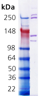
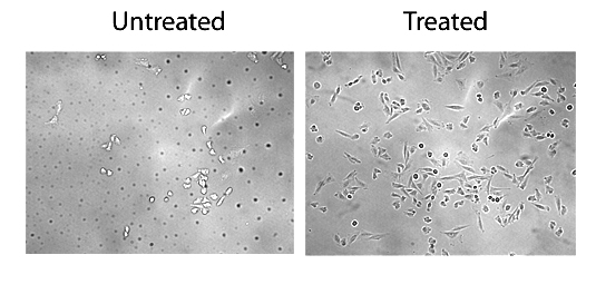
Glass coverslips were coated with or without collagen in 60% ethanol and allowed to dry overnight. CHO cells were plated at onto the coverslips and grown for two days at 37oC with 5% CO2. Cells were fixed in 10% buffered formalin. Coverslips were washed in PBS, mounted upside down onto microscope slides, and imaged with a 20X objective.


Product Details
Activity |
Tested in cell proliferation assay – Increased attachment of cells on collagen coated coverslips. |
---|---|
Alternative Name |
COL1 |
Appearance |
Hazy viscous liquid. |
Application Notes |
Can be used for preparation of collagen gels, thin layer coating of surfaces (culture plates, slides, coverslips). |
Concentration |
5mg/ml |
Endotoxin Content |
<100 EU/mg purified protein (LAL test) |
Formulation |
Sterile liquid. In 0.02N acetic acid. |
MW |
235kDa, 215kDa, 130kDa, 115kDa |
Purity |
≥90% (SDS-PAGE) |
Source |
Isolated from rat tail tendons. |
Handling & Storage
Use/Stability |
As indicated on product label or CoA when stored as recommended. |
---|---|
Handling |
Keep sterile. Do not freeze. |
Short Term Storage |
+4°C |
Long Term Storage |
+4°C |
Shipping |
Blue Ice |
Regulatory Status |
RUO – Research Use Only |
---|
- Glypican 3 as target therapy to prevent cell migration and proliferation in rhabdomyosarcoma: Bacchiega, M., D’Agostino, S., et al.; Sci. Rep. 15, 20913 (2025), Abstract
- Glypican 3 as target therapy to prevent cell migration and proliferation in rhabdomyosarcoma: Bacchiega, M., D’Agostino, S., et al.; Research Square , (2025)
- A human model to deconvolve genotype-phenotype causations in lung squamous cell carcinoma: Ogden, J., Sellers, R., et al.; Nat. Commun. 16, 3215 (2025), Abstract
- Optogenetically engineered Septin-7 enhances immune cell infiltration of tumor spheroids: Chen, J., Hnath, B., et al.; PNAS 121, e2405717121 (2024), Abstract
- Five near-infrared-emissive graphene quantum dots for multiplex bioimaging: Valimukhametova, A., Fannon, O., et al.; 2d Mater. 11, (2024), Abstract
- Doped Graphene Quantum Dots as Biocompatible Radical Scavenging Agents: Bhaloo, A., Nguyen, S., et al.; Antioxidants (Basel) 12, (2023), Abstract
- A novel human model to deconvolve cell-intrinsic phenotypes of genetically dysregulated pathways in lung squamous cell carcinoma: Ogden, J., Sellers, R., et al.; bioRxiv , (2023)
- Modeling Gas Plasma-Tissue Interactions in 3D Collagen-Based Hydrogel Cancer Cell Cultures: L. Miebach, et al.; Bioengineering (Basel) 10, 367 (2023), Abstract
- Dia1 coordinates differentiation and cell sorting in a stratified epithelium: Harmon, R. M., Devany, J., et al.; J. Cell Biol. 221, (2022), Abstract
- Dual-Mode Fluorescence/Ultrasound Imaging with Biocompatible Metal-Doped Graphene Quantum Dots: Valimukhametova, A. R., Zub, O. S., et al.; ACS Biomater. Sci. Eng. 8, 4965 (2022), Abstract
- Differential gene expression response of synovial fibroblasts from temporomandibular joints and knee joints to dynamic tensile stress: Nazet, U., Neubert, P., et al.; J. Orofac. Orthop. 83, 361 (2022), Abstract
- A TNF receptor 2 agonist ameliorates neuropathology and improves cognition in an Alzheimer’s disease mouse model: N.O. Casan, et al.; PNAS 119, e2201137119 (2022), Abstract
- Efficacy of probiotic Streptococcus thermophilus in counteracting TGF-β1-induced fibrotic response in normal human dermal fibroblasts: F. Lombardi, et al.; J. Inflamm. 19, 27 (2022), Abstract
- Cooperative interaction between ERα and the EMT-inducer ZEB1 reprograms breast cancer cells for bone metastasis: N.M. Ghahhari, et al.; Nat. Commun. 13, 2104 (2022), Abstract
- SIRT1-mediated deacetylation of FOXO3a transcription factor supports pro-angiogenic activity of interferon-deficient tumor-associated neutrophils: S. Bordbari, et al.; Int. J. Cancer 150, 1198 (2022), Abstract
- Axon guidance receptor ROBO3 modulates subtype identity and prognosis via AXL-associated inflammatory network in pancreatic cancer: N. Krebs, et al.; JCI Insight 7, e154475 (2022), Abstract
- Low nanogel stiffness favors nanogel transcytosis across an in vitro blood-brain barrier: L. Ribovski, et al.; Nanomedicine 34, 102377 (2021), Abstract
- Generation and Encapsulation of Human iPSC-Derived Vascular Smooth Muscle Cells for Proangiogenic Therapy: Dash, B. C., Hsia, H. C., et al.; Methods Mol. Biol. 2549, 259 (2021), Abstract
- Mechanical Stress Induce PG-E2 in Murine Synovial Fibroblasts Originating from the Temporomandibular Joint: Nazet, U., Feulner, L., et al.; Cells 10, (2021), Abstract
- Integrin β3 targeting biomaterial preferentially promotes secretion of bFGF and viability of iPSC-derived vascular smooth muscle cells: B. C. Dash, et al.; Biomater. Sci. 9, 5319 (2021), Abstract
- A new perfusion culture method with a self-organized capillary network: K. Sugihara, et al.; PLoS One 15, e0240552 (2020), Abstract — Full Text
- Induced pluripotent stem cell-derived smooth muscle cells increase angiogenesis and accelerate diabetic wound healing: J. Gorecka, et al.; Regen. Med. 15, 1277 (2020), Abstract — Full Text
- Increased Lamin B1 Levels Promote Cell Migration by Altering Perinuclear Actin Organization: Fracchia, A., Asraf, T., et al.; Cells 9, (2020), Abstract
- The anti-inflammatory peptide Catestatin blocks chemotaxis: Muntjewerff, E. M., Parv, K., et al.; bioRxiv , (2020)
- A filter-free blood-brain barrier model to quantitatively study transendothelial delivery of nanoparticles by fluorescence spectroscopy: E. De Jong, et al.; J. Control. Release 289, 14 (2018), Abstract
- Effects of selexipag and its active metabolite in contrasting the profibrotic myofibroblast activity in cultured scleroderma skin fibroblasts: M. Cutolo, et al.; Arthritis Res. Ther. 20, 77 (2018), Abstract — Full Text
Datasheet, Manuals, SDS & CofA
Manuals And Inserts
Specific Protocol
Firm Gelling Procedure
(Note: The ideal concentration for gelling is 1 - 5mg/ml. The end user will need to determine the appropriate concentration for their specific purpose).
Ammonium Hydroxide Gas Method
- Dilute 5mg/ml collagen to the desired concentration using sterile conditions. (Note: Lower concentrations will have reduced rigidity).
- Add enough collagen to cover the surface.
- Saturate a cotton ball or piece of filter paper with concentrated ammonium hydroxide and make a closed vapor chamber.
- Heat to 37°C until gel forms.
- Remove the ammonium hydroxide.
- Soak the gel surface in PBS for 1 hour.
- Wash several times PBS.
- Store the gel in PBS at +4°C until use.
- Equilibrate the gel in the desired media for 30 minutes prior to use.
NaOH Method (For 10ml)
- Sterilely add 1- 7ml of 5mg/ml collagen to a tube depending on desired final concentration. (Note: Lower concentrations will have reduced rigidity).
- Add 1ml of sterile 10X PBS.
- Add 1 ml of sterile 1N NaOH.
- Bring final volume to 10ml with sterile dH2O
- Cover the desired surface with a layer of the collagen and place at 37°C until the gel has solidified. Gel will only form if the pH is ≥7.0.
- Store in PBS at +4°C until use.
- Equilibrate the gel in the desired media for 30 minutes prior to use.
Certificate of Analysis
Please enter the lot number as featured on the product label
SDS
Enzo Life Science provides GHS Compliant SDS
If your language is not available please fill out the SDS request form