 Lab Essentials
 AMPIVIEW® RNA probes
 Enabling Your Projects
  GMP Services
 Bulk Solutions
 Research Travel Grant
 Have You Published Using an Enzo Product?
Bradykinin (BK) is a nine-amino acid peptide produced by kallikrein cleavage of kininogen precursor proteins in tissue and plasma, and is an important regulator of vascular and pain responses to tissue injury. Two receptors (B1 and B2) have been identified as mediators of BK signaling, and are both members of the seven-transmembrane domain-containing G-protein coupled receptor (GPCR) family. The B2 receptor is constitutively expressed in smooth muscle and neurons, whereas B1 expression is induced following tissue injury or during inflammation. The B2 receptor displays high affinity for BK and Lys-BK peptide agonists, while the B1 receptor displays highest affinity to des-Arg9-BK and des-Arg10-kallidin. Both receptors are best characterized as signaling via coupling to Gq alpha subunits (particularly Gq/11), leading to activation of phospholipase C beta, hydrolysis of PI, and an intracellular increase in free calcium, although coupling through Gi, Gs, and G12/13 has also been observed.
Shipping: Available products typically ship within 24/48h, via priority shipping.
Do you need support? Contact Customer Service or Technical Support.
Online Account
Access or Create Your Account
This antibody is covered by our Worry-Free Guarantee.

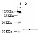
Western blot analysis: MW marker (1) and 25µg rat heart extract (2) probed with Bradykinin Receptor (B1) pAb at 0.75µg/ml.
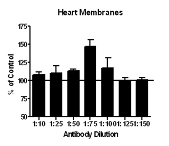
Membrane ELISA: Lewis rat heart membranes (5µg/well) were treated with 1 μM concentrations of agonist (BK-1-8) and probed with Bradykinin Receptor (B1) pAb (1:10 to 1:150 of a 0.1µg/μl stock solution) by ELISA. Data from vehicle treated cells were taken as 100%. Results are the mean ± SEM (n=6).
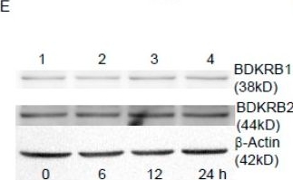
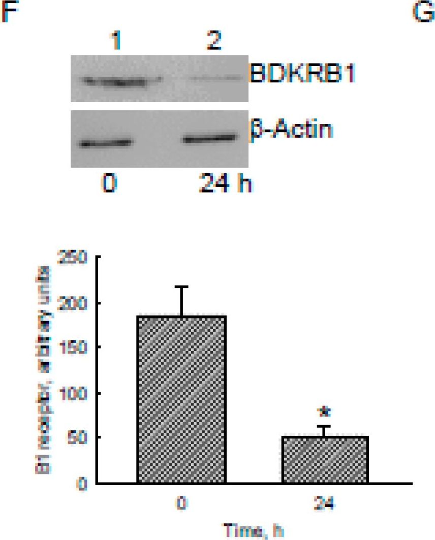
Effects of bradykinin on viability, levels, and functions of bradykinin receptor (BDKR) B1/2 in human malignant glioblastoma cells. Human U87 MG glioblastoma cells were stained with a fluorescent 4’,6-diamidino-2-phenylindole (DAPI) dye and reacted with a monoclonal antibody against glial fibrillary acidic protein (GFAP), a biomarker of astrocytes (A). Fluorescent signals were observed and analyzed using confocal microscopy. U87 MG cells were treated with 100 nM bradykinin for 6, 12, and 24 h or with 10, 50, and 100 nM bradykinin for 24 h. Cell morphologies were observed and photographed using a light microscope (B). Cell survival was analyzed using a trypan blue exclusion method (C,D). Levels of BDKRB1 and BDKRB2 were immunodetected (E, top two panels). β-Actin was analyzed as an internal control (bottom panel). These protein bands were quantified and statistically analyzed (F). After exposure to bradykinin and Fluo3, dynamic changes in levels of intracellular calcium (Ca2+) were immediately observed and recorded by confocal microscopy (G). Marked enhancement of fluorescent signals showed the increased intensities of intracellular Ca2+ following bradykinin treatment (H). Each value represents the mean ± standard deviation (SD) for n = 9. Representative immunoblots and confocal images are shown. An asterisk (*) indicates that a value significantly (p < 0.05) differed from the respective control. Scale bar, 20 μm.
Image collected and cropped by CiteAb under a CC-BY license from the following publication: The Bradykinin-BDKRB1 Axis Regulates Aquaporin 4 Gene Expression and Consequential Migration and Invasion of Malignant Glioblastoma Cells via a Ca2+-MEK1-ERK1/2-NF-κB Mechanism. Cancers (Basel) (2020)

Effects of bradykinin on aquaporin-4 (AQP4) mRNA and protein expressions in human malignant glioblastoma cells. Human U87 MG glioblastoma cells were treated with 100 nM bradykinin for 3, 6, 12, and 24 h. Levels of AQP4 mRNA were analyzed using an RT-PCR (A, top panel). Amounts of β-actin mRNA were examined as an internal control (bottom panel). These DNA bands were quantified and statistically analyzed (B). Expression of AQP4 mRNA was further quantified using a real-time polymerase chain reaction (PCR) analysis (C). Human U87 MG cells were exposed to bradykinin for 24 h. Levels and distribution of AQP4 were immunodetected (D, left panel). The nucleus was stained with 4’,6-diamidino-2-phenylindole (DAPI) (middle panel). The merged signals are shown in the right panel (D) and were quantified and statistically analyzed (E). Expression of the bradykinin receptor (BDKR) B1 was knocked-down using RNA interference. Control cells received scrambled siRNA. Levels of BDKRB1 were immunodetected (F, top panel). β-Actin was immunodetected as an internal control. These protein bands were quantified and statistically analyzed (bottom panel). Human U87 MG cells were pretreated with BDKRB1 siRNA and then exposed to bradykinin. Expression of AQP4 mRNA was quantified with a real-time PCR (G). Each value represents the mean ± standard deviation (SD), n = 9. Symbols * and # indicate that the values significantly (p < 0.05) differed from the control and BDKRB1 siRNA-treated groups, respectively. Representative DNA agarose gels, confocal images, and immunoblots are shown. Scale bar, 5 μm.
Image collected and cropped by CiteAb under a CC-BY license from the following publication: The Bradykinin-BDKRB1 Axis Regulates Aquaporin 4 Gene Expression and Consequential Migration and Invasion of Malignant Glioblastoma Cells via a Ca2+-MEK1-ERK1/2-NF-κB Mechanism. Cancers (Basel) (2020)


| Regulatory Status |
RUO – Research Use Only |
|---|
Related Products
| Application | ELISA, IHC, WB |
|---|---|
| Host | Goat |
| Species Reactivity | Rabbit |
Last modified: May 29, 2024