 Lab Essentials
 AMPIVIEW® RNA probes
 Enabling Your Projects
  GMP Services
 Bulk Solutions
 Research Travel Grant
 Have You Published Using an Enzo Product?
Nucleic Acid Detection

The Power of in situ Hybridization (ISH) in Spatial Biology
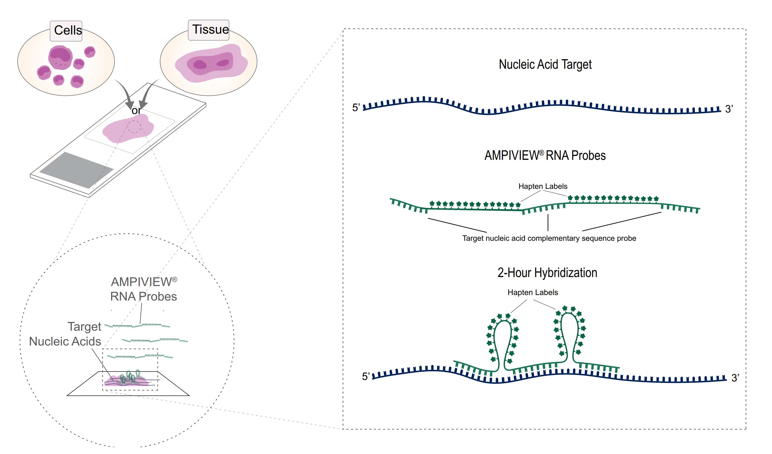
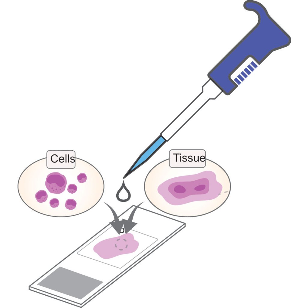
Enzo’s spatial nucleic acid detection platform empowers researchers to visualize gene expression and genomic alterations within the native architecture of cells and tissues.
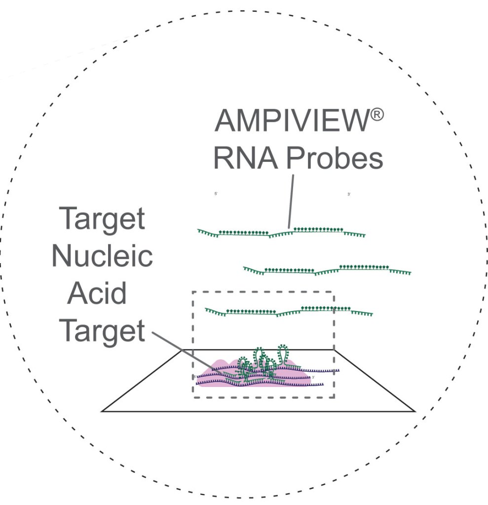
At the core of this offering are our AMPIVIEW® RNA Probes, powered by Enzo’s LoopRNA™ ISH technology to deliver superior sensitivity, flexibility and adaptability. This is a powerful, probe-based in situ hybridization system designed to detect specific DNA, DNA/RNA or RNA transcripts at the single-cell level with exceptional clarity and specificity.
Versatile Applications
Detection of unique nucleic acid targets in FFPE tissues and cells with mild protocols to preserve morphology
Flexible and Adaptable Workflow
Ready for both manual and automated workflows, ensuring compatibility with various research setups
Spatial Biology Insights
Enable detailed spatial analysis, allowing researchers to visualize and understand the spatial context of gene expression and interactions within tissues
Uniquely Designed AMPIVIEW® RNA Probes
Powered by Enzo’s LoopRNA™ ISH Technology
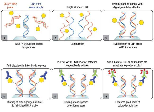
Hapten Labels
Biotin
DNP
Digoxigenin
Fluorescein
AMPIVIEW® RNA Probes Workflow
Step 1 : Pre-Treatment
Step 2 : 2-Hour Hybridization
Step 3 : Amplification and Detection
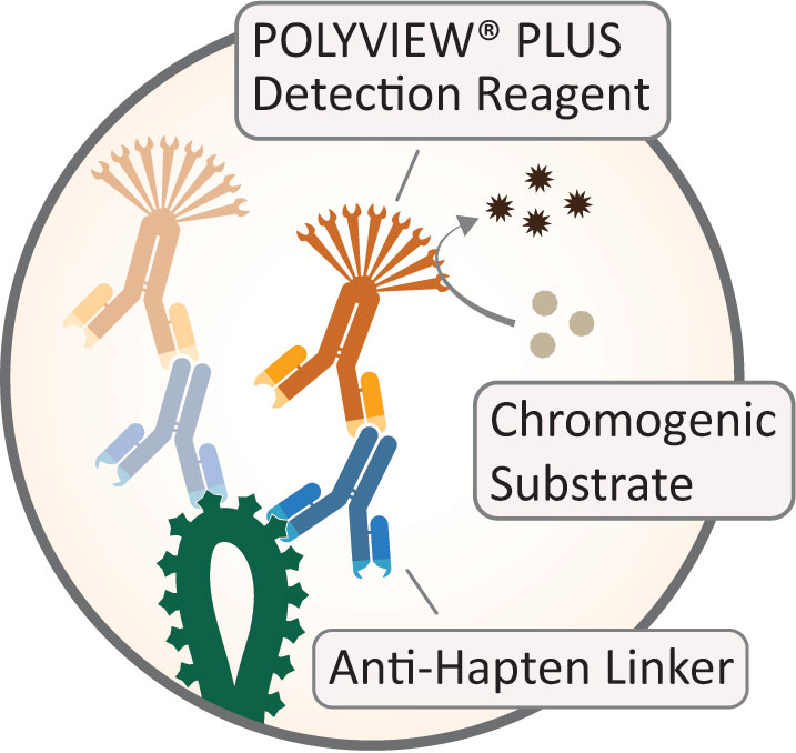
Chromogen Detection
Biotin-conjugated Probes
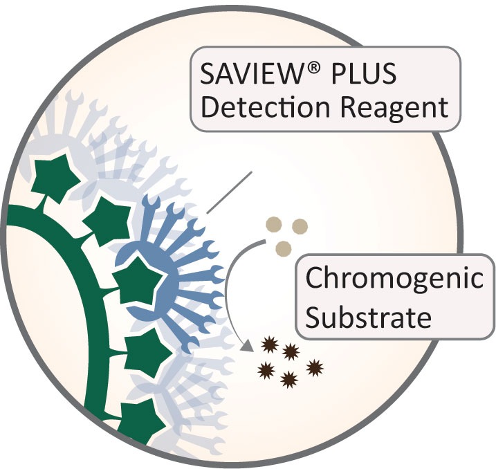
Hapten-conjugated Probes
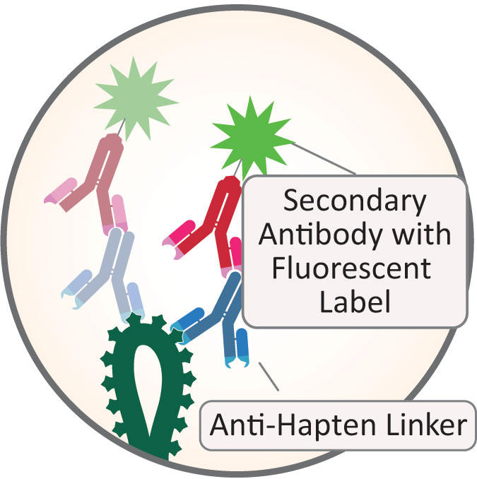
Fluorescence Detection
Hapten-conjugated Probes
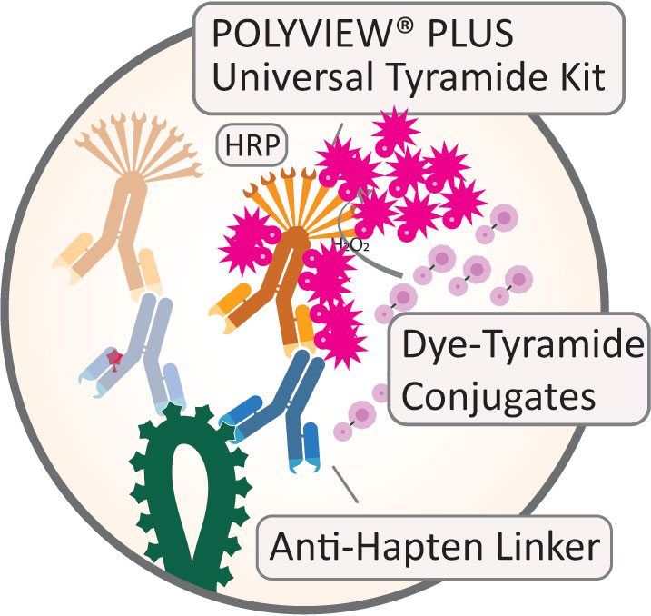
Hapten-conjugated Probes
Step 4 : Visualization
Light Microscope
Fluorescent Microscope
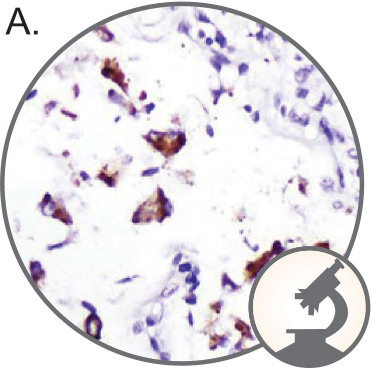
A. SARS-CoV-2 (brown) detected in COVID-19 infected lung tissue with AMPIVIEW® SARS-CoV-2 RNA Probes Set (ENZ-GEN159), detected with SAVIEW® PLUS HRP reagent combined with HIGHDEF® DAB Chromogen/Substrate (ENZ-ACC105) and counterstained with HIGHDEF® Hematoxylin (ENZ-ACC106).
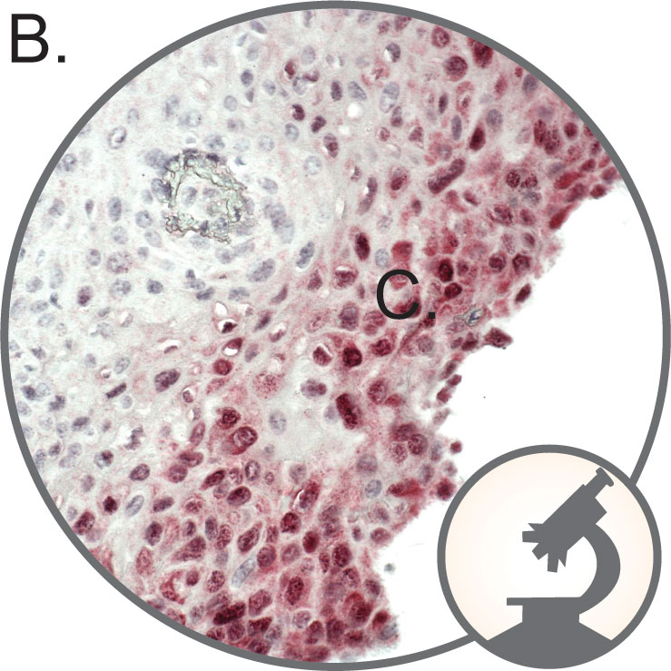
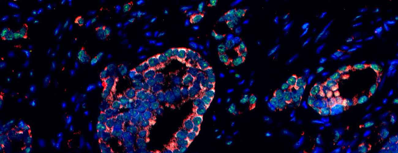
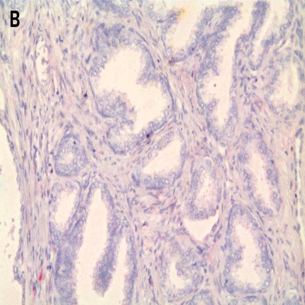
B. HPV 16, 18, 31 ,33 or 51 (red) detected in cervical cancer tissue with AMPIVIEW® HPV High-Risk RNA Probes Set (ENZ-GEN148), amplified with DIGX® Rabbit Anti-Digoxigenin Linker (ENZ-ABS303), detected with POLYVIEW® PLUS AP (anti-rabbit) (ENZ-ACC110), combined with HIGHDEF® Red IHC Chromogen (AP) (ADI-950-140) and counterstained with HIGHDEF® Hematoxylin (ENZ-ACC106)
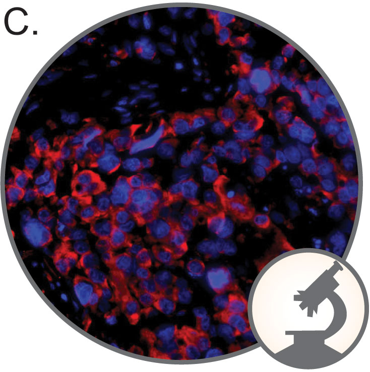
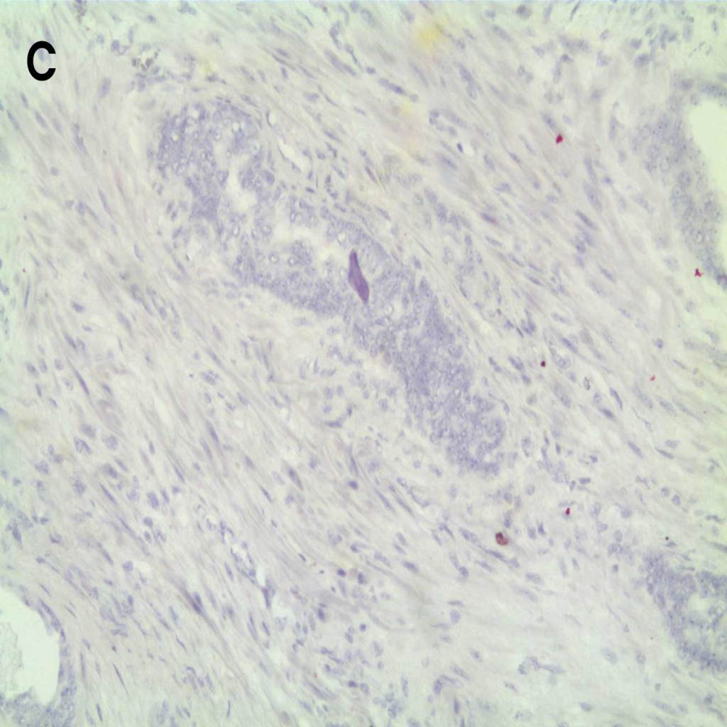
C. HER-2 (red) detected in breast cancer tissue with AMPIVIEW® HER-2 (AS) Dig RNA Probes Set (ENZ-GEN139), amplified with DIGX® Rabbit Anti-Digoxigenin linker (ENZ-ACC303), detected with POLYVIEW® PLUS Universal Tyramide Kit and counterstained with DAPI.
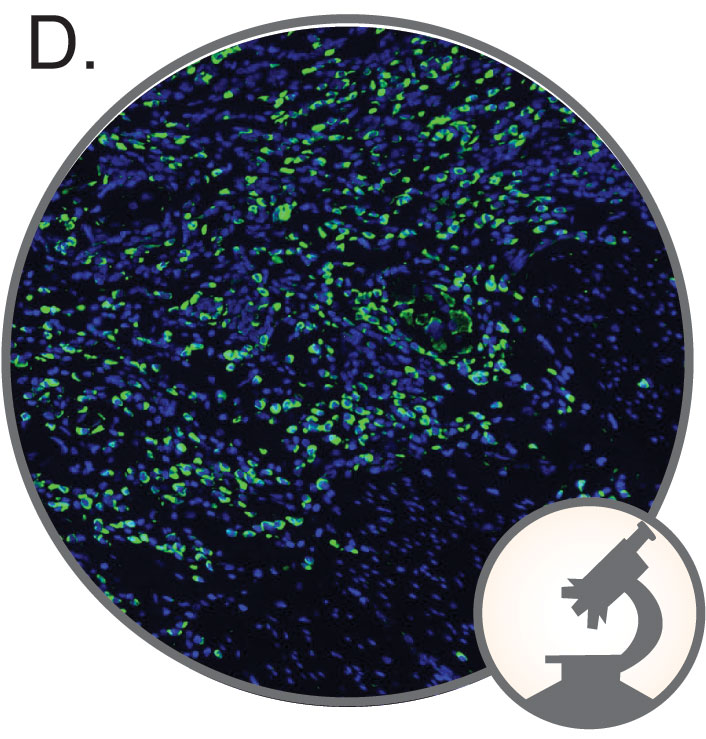
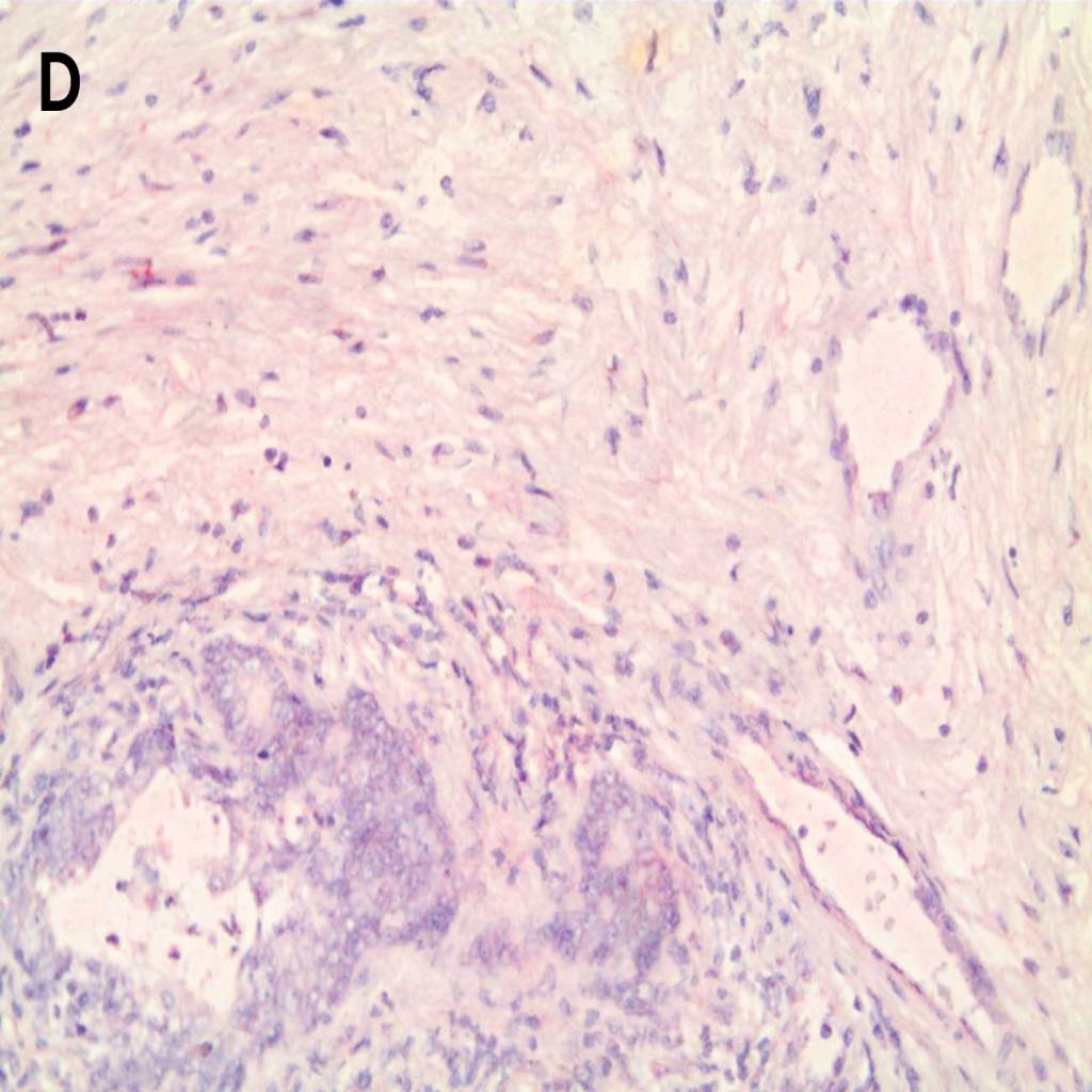
D. PD-1 (green) detected in lung cancer tissue with AMPIVIEW® PD-1 (AS) Dig RNA Probes Set (ENZ-GEN281), amplified with DIGX® Rabbit Anti-Digoxigenin linker (ENZ-ACC303), detected with anti-rabbit secondary antibodies conjugated with fluorescent label and counterstained with DAPI.
Singleplex Detection of HER-2 in Breast Cancer Tissue
HER-2 detected in breast cancer tissue with A.-B. AMPIVIEW® HER-2 (AS) Dig RNA Probes and C.-D. with AMPIVIEW® NSP Dig RNA Probes (negative control probe). AMPIVIEW® RNA probes have been amplified with DIGX® rabbit anti-digoxigenin linker and detected with POLYVIEW® PLUS AP (anti-rabbit) reagent, combined with HIGHDEF® Red AP Chromogen Kit and counterstained with HIGHDEF® Hematoxylin.
Singleplex Detection of PD-1 in Ovarian and Prostate Cancer Tissues
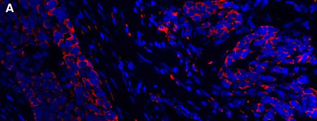
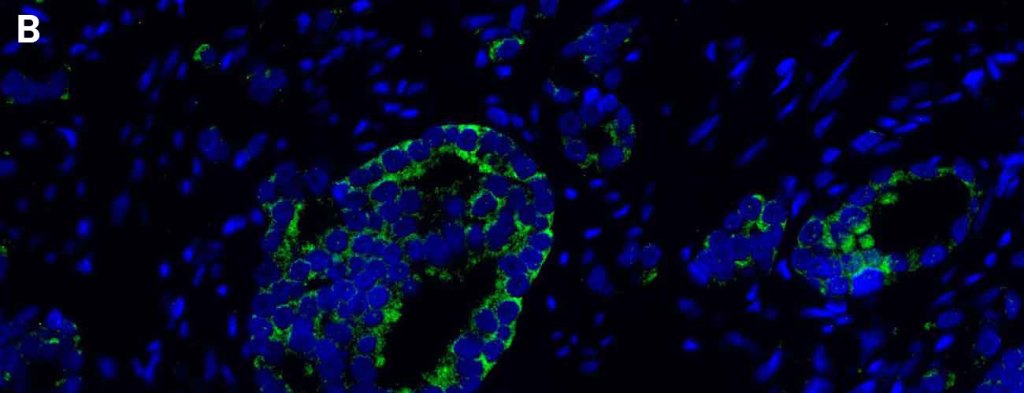
PD-1 was fluorescently detected with AMPIVIEW® PD-1 (AS) Dig RNA Probes in A. ovarian cancer tissue (red signal) and B. prostate cancer tissue (green signal). DAPI was used as a nuclear marker.
Multiplex Nucleic Acid Targets in Different Types of FFPE Tissues
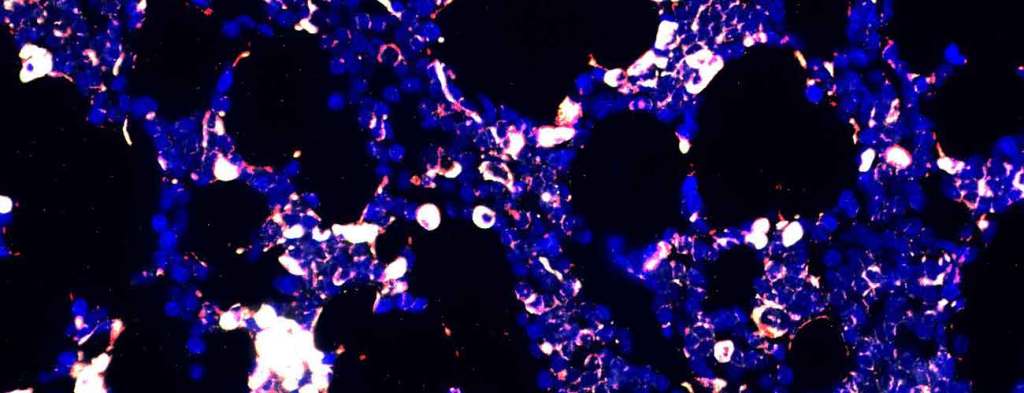
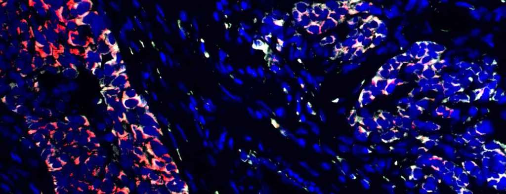
PD-1 (grey), PD-L1 (red) and EGFR (green) detected in A. Mantle lymphoma tissue, B. ovarian cancer tissue and C. prostate cancer tissue with AMPIVIEW® PD-1 (AS) Dig RNA Probes, AMPIVIEW® PD-L1 (AS) DNP RNA Probes and AMPIVIEW® EGFR (AS) Dig RNA Probes. DAPI (blue) was used as a nuclear marker
Multiplex Detection of Non-Coding RNAs and mRNAs in Brain Tissue
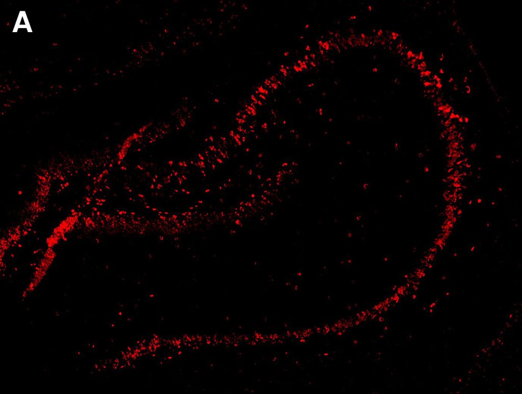
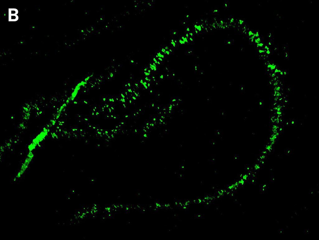
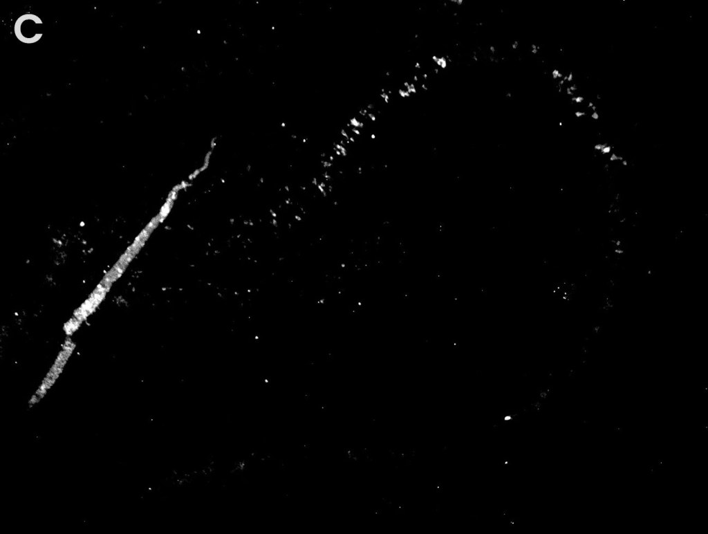
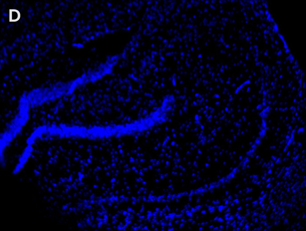
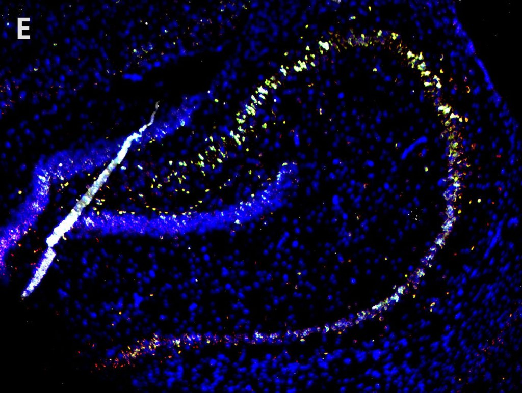
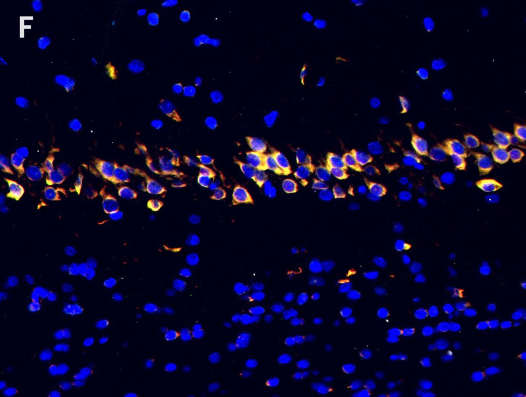
Detection of A. Nestin RNA (red), B. non-coding RNA activated by DNA damage (Norad) (green), C. circular RNA Circ018 (gray) and nuclear marker, DAPI, in rat brain tissue by hybridizing AMPIVIEW® RNA probes against Nestin, Norad and Circ018. Co-localization of Nestin and Norad indicates that the majority of the neural stem cells in the hippocampus express lncRNA Norad, but only a subset in the CA3 regions express Circ018. E. 4X magnification, F. 20X magnification.
AMPIVIEW® RNA Probes Achieve PCR-Level Single-Copy Sensitivity While Preserving Crucial Tissue Morphology
Single cell resolution further expands the understanding of gene expression in cell lines and tissue samples.
AMPIVIEW® RNA probes patented design enables highly specific and sensitive detection of target RNA, DNA or DNA/RNA, delivering robust, high-signal-to-noise performance at the single-molecule level with single-cell resolution. Powered by Enzo’s LoopRNA™ ISH technology, this powerful tool allows precise identification of genes and gene transcripts, deepening insights into gene expression, spatial localization within its morphological context.
High Sensitivity and Reliability
Superior sensitivity for detecting unique nucleic acid targets (DNA/RNA) down to a single cell level
Preserved Morphology
Mild protocols ensure that tissue morphology is maintained while providing clear visualization under the light microscope with chromogenic detection or fluorescence microscope with fluorescence detection
Customizable
Probes can be labeled with biotin, Dig, DNP, or FITC, enabling precise targeting of any gene across any species
Cost-Effective
Optimized with Enzo’s cost-effective IHC/ISH detection portfolio, making them a budget-friendly option for researchers
Quantification of RNA at a Single-cell Resolution
| Count (Average per cell) | |
|---|---|
| ISH Sample 1 | 15 |
| ISH Sample 2 | 13 |
| PCR | 11/cell |
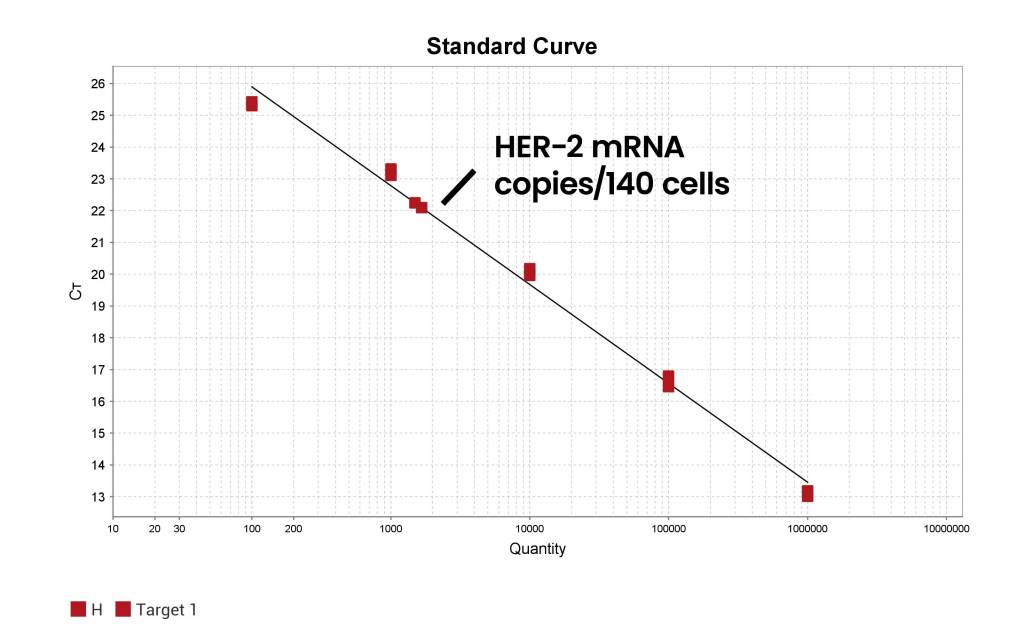
| Cell Line | Total mRNA/cell | mRNA/cell |
|---|---|---|
| MCF10 | 1578/140cells | 11.3/cell |
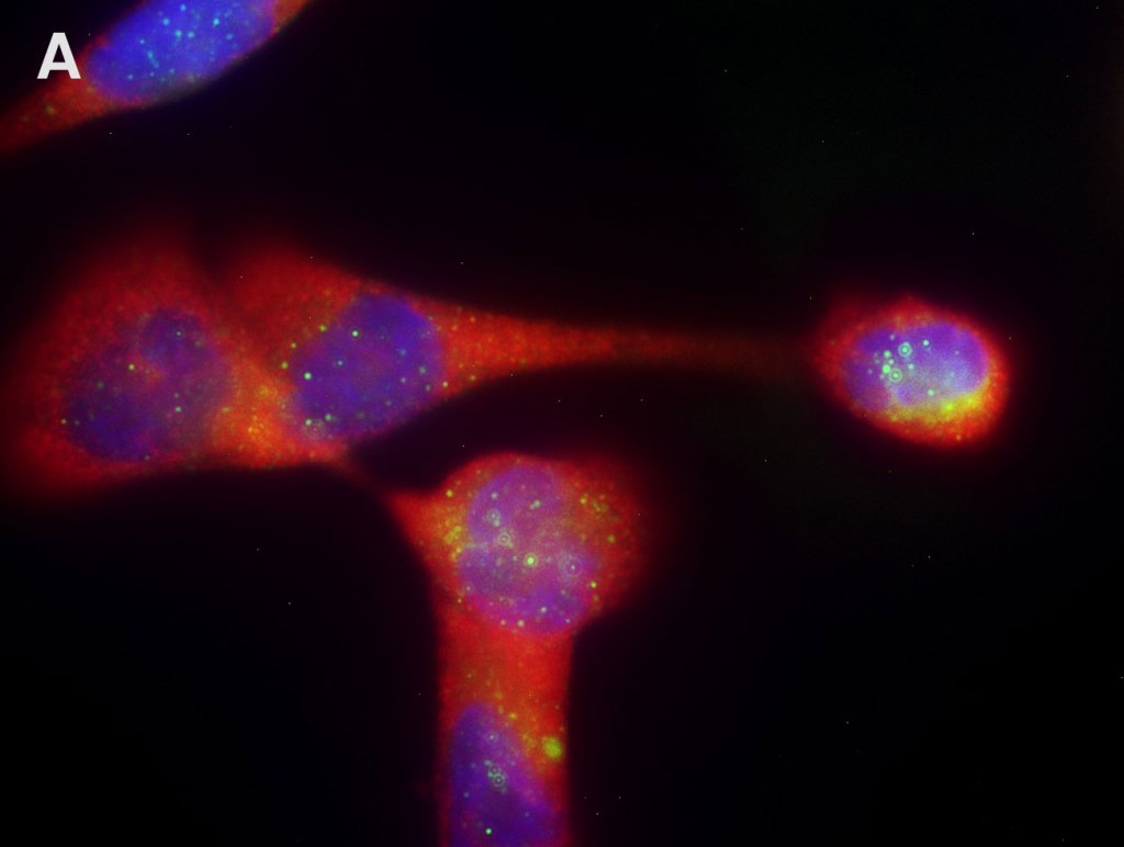
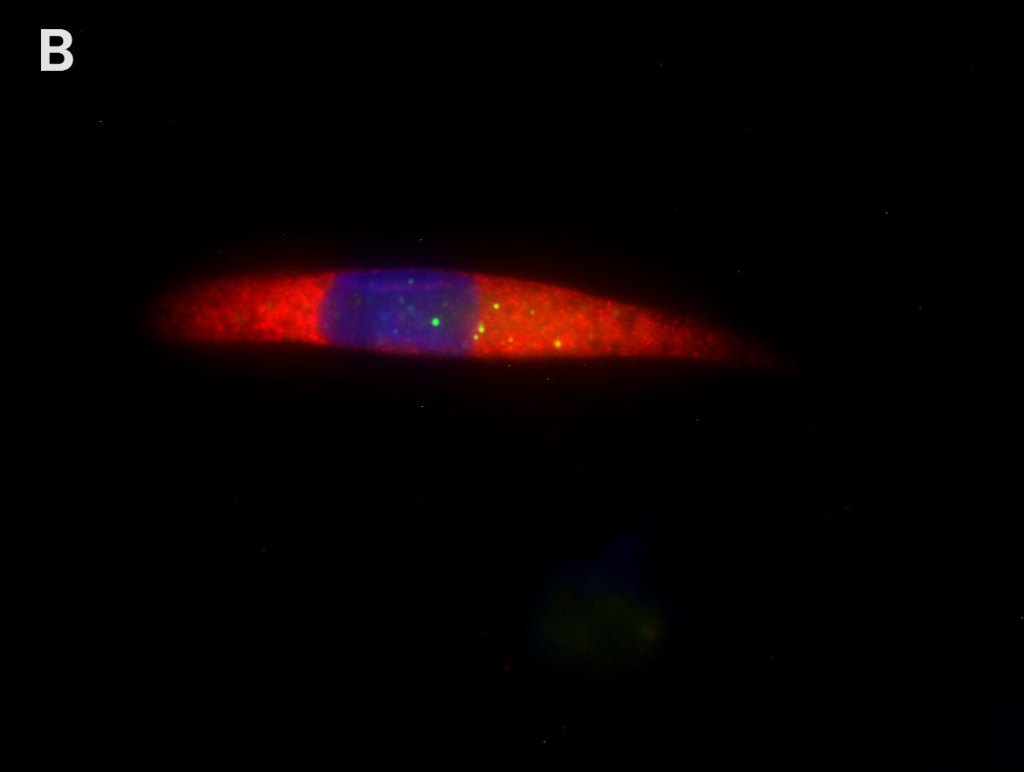
A. B. HER-2 mRNA (green) was visualized with AMPIVIEW® HER-2 (AS) Dig RNA Probes Set (ENZ-GEN139) and quantified by counting green signal dots in individual MCF10A cells. Nuclei were counterstained with DAPI (blue) and AMPIVIEW® 18S rRNA Probes was used as internal control for RNA detection (red). C. HER-2 mRNA in the same MCF10A cell culture was quantified using absolute PCR quantification method (according to manufacturer’s protocol) to estimate the copy number of HER-2 mRNA in single cells. The estimates from both methods were in close agreement, demonstrating the sensitivity and the quantification capability of AMPIVIEW® RNA probes.
Webinar
Quantifying Targeted mRNA in Single Cells via In Situ Hybridization with Enzo AMPIVIEW®RNA Probes
AMPIVIEW® RNA Probes Outperforms the Competition with Superior Signal Clarity and Minimal Background
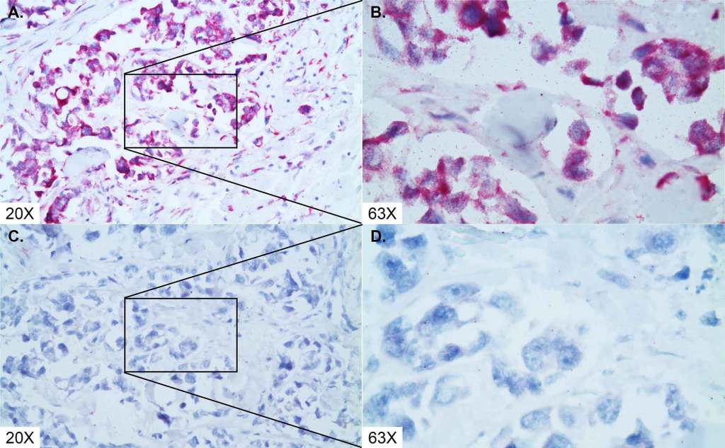
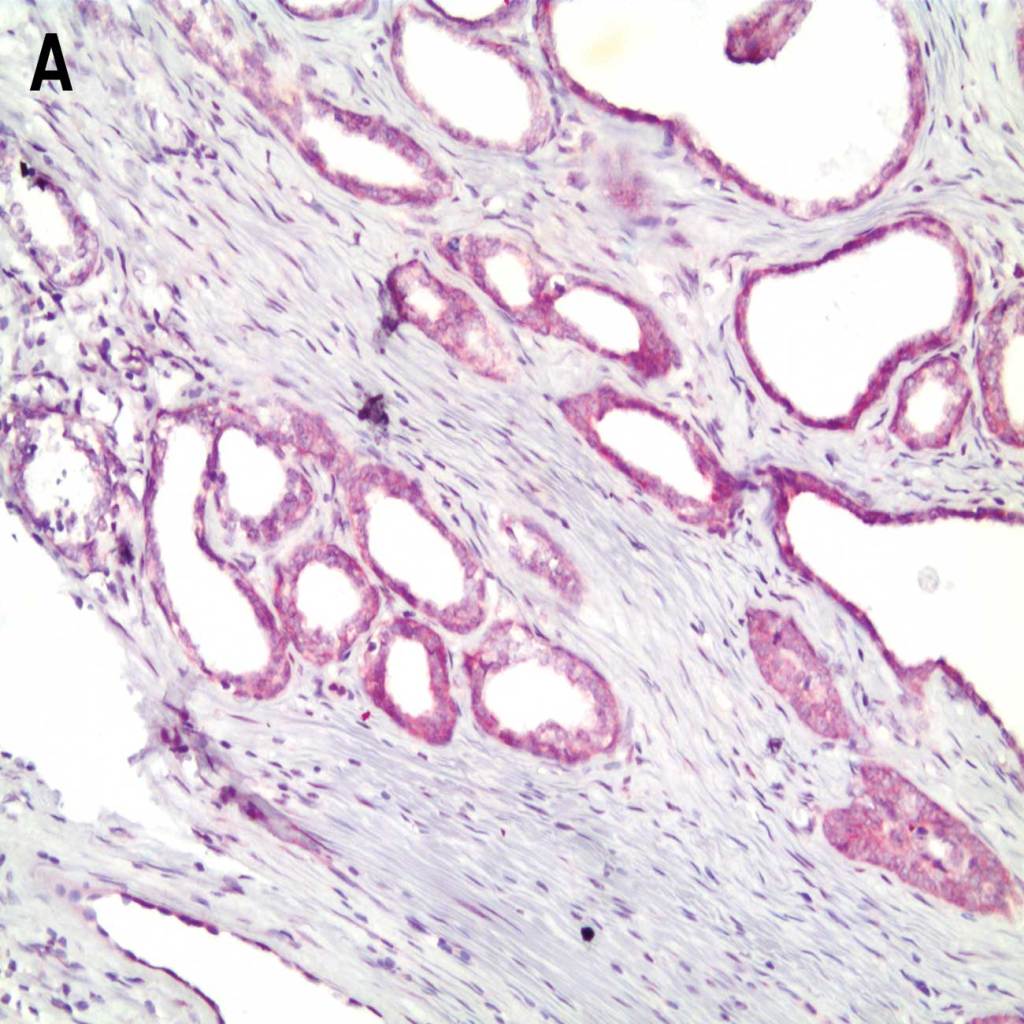
Reliable Detection of Ubiquitin as Positive Control in Prostate Cancer Tissue
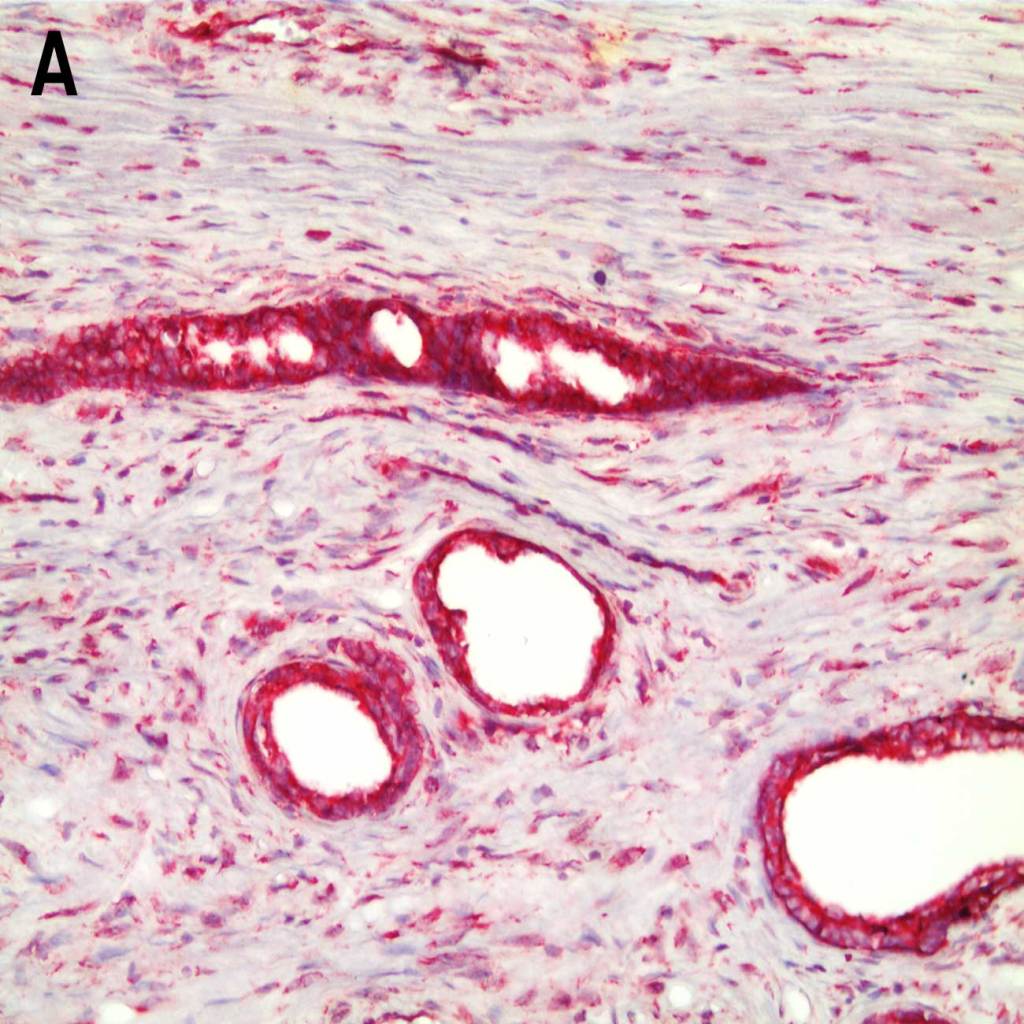
Detection of ubiquitin in prostate cancer tissue with A. C. AMPIVIEW® Ubiquitin (AS) Dig RNA probes (ENZ-GEN125) and B. D. Leading competitor’s probes. ISH and target detection performed according to manufacturer’s protocol.
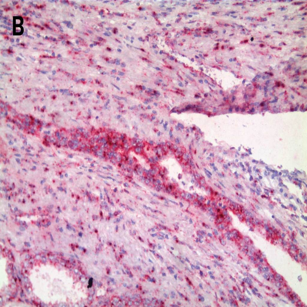
Sharper Signal for EGFR and Cleaner Background with AMPIVIEW® RNA Probes in Prostate Tissue
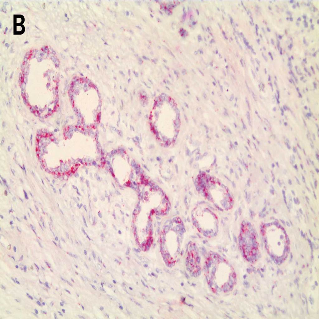
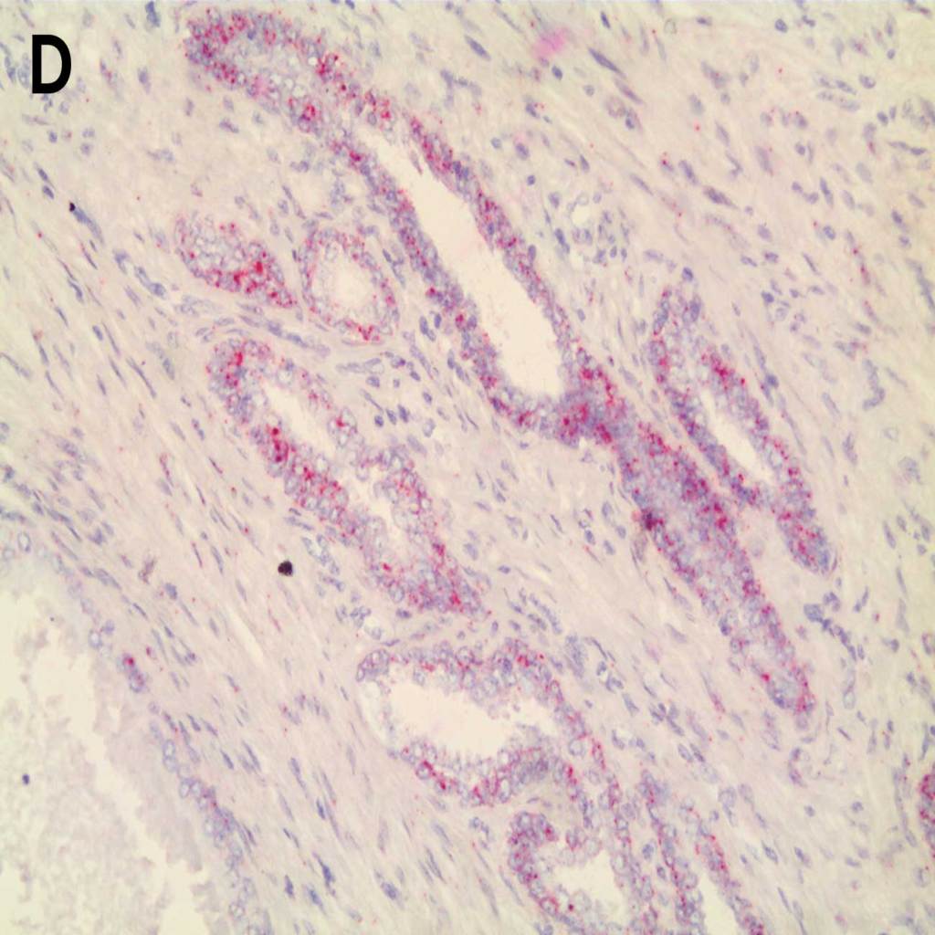
Detection of EGFR in prostate cancer tissue with A. C. AMPIVIEW® EGFR (AS) Dig RNA probes (ENZ-GEN129) and B. D. Leading competitor’s probes. ISH and target detection performed according to manufacturer’s protocol.
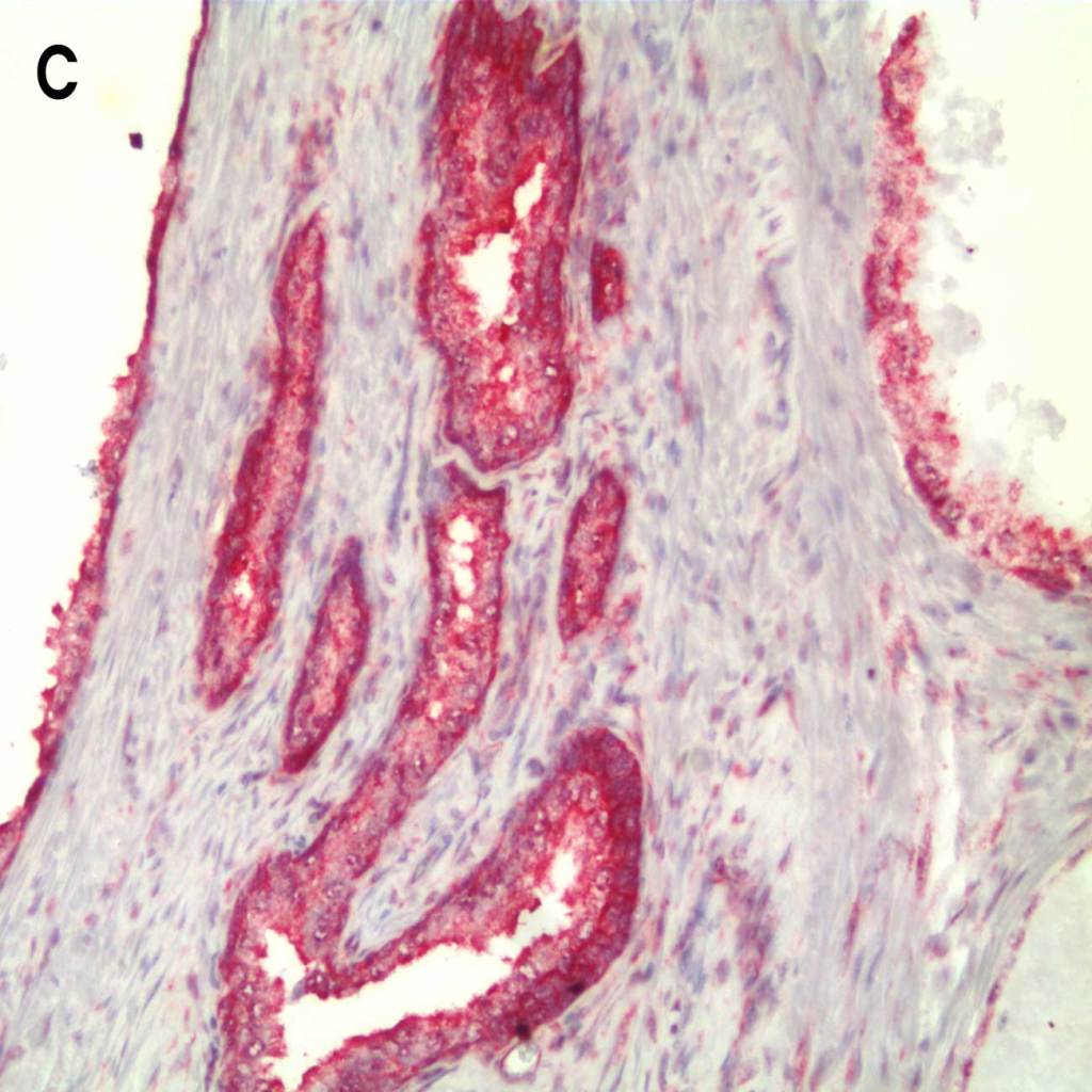
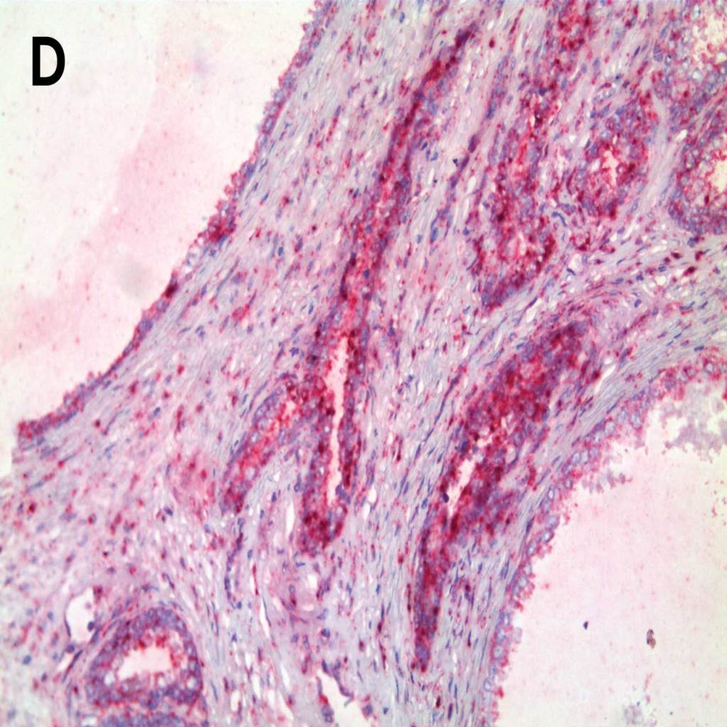
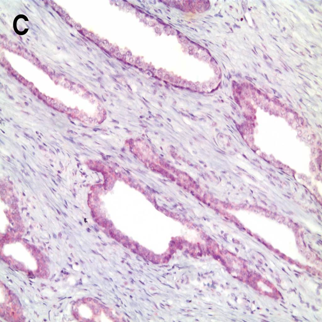
Clean Background Matters: Negative Control Comparison of AMPIVIEW® RNA Probes vs Competition

Comparison of negative control probes in prostate cancer tissue with A. C. AMPIVIEW® NSP Dig RNA Probes and B. D. leading competitor’s negative control probes.
Featured AMPIVIEW® RNA Probes
Cancer Targets
AMPIVIEW® HER-2 (AS) Dig RNA Probes Set
AMPIVIEW® HER-2 (AS) DNP RNA Probes Set
AMPIVIEW® GATA3 (AS) Dig RNA Probes Set
AMPIVIEW® CD19 (AS) Dig RNA Probes Set
AMPIVIEW® Bcl2 (AS) Dig RNA Probes Set
AMPIVIEW® PD-1 (AS) Dig RNA Probes Set
AMPIVIEW® PTEN (AS) DNP RNA Probes Set
AMPIVIEW® Ki67 (AS) Dig RNA Probes Set
AMPIVIEW® Ki67 (AS) DNP RNA Probes Set
AMPIVIEW® p53 (AS) Dig RNA Probes Set
HPV Targets
AMPIVIEW® HPV 6/11 (AS) Dig RNA Probes Set
AMPIVIEW® HPV 16/18 (AS) Dig RNA Probes Set
AMPIVIEW® HPV 31/33/51 (AS) Dig RNA Probes Set
AMPIVIEW® HPV High Risk (AS) Dig RNA Probes Set
AMPIVIEW® HPV HR-15 (AS) DNP RNA Probes Set
Neuroscience Targets
AMPIVIEW® Nestin (AS) Dig RNA Probes (Rat) Set
AMPIVIEW® Wnt5a (AS) Dig RNA Probes (Rat) Set
AMPIVIEW® Wnt5a (AS) DNP RNA Probes Set
AMPIVIEW® NORAD (AS) Dig RNA Probes (Mouse) Set
AMPIVIEW® Circ018 (AS) Dig RNA Probes (Mouse) Set
Infectious Diseases Targets
AMPIVIEW® SARS-CoV-2 Spike (AS) Bio RNA Probes Set
AMPIVIEW® SARS-CoV-2 Nucleocapsid (AS) Bio RNA Probes Set
AMPIVIEW® SARS-CoV-2 RNA Probes Set
Detection Kits
AMPIVIEW® Dig (Anti-Mouse) Detection HRP-DAB Kit
AMPIVIEW® Dig (Anti-Rabbit) Detection AP-Red Kit
AMPIVIEW® Dig (Anti-Rabbit) Detection HRP-DAB Kit
AMPIVIEW® Dig (Anti-Mouse) Detection AP-Red Kit
AMPIVIEW® DNP (Anti-Rabbit) Detection AP-Red Kit
AMPIVIEW® DNP (Anti-Rabbit) Detection HRP-DAB Kit
Controls
AMPIVIEW® Ubiquitin (AS) Dig RNA Probes Set
AMPIVIEW® GAPDH (AS) Dig RNA Probes Set