 Lab Essentials
 AMPIVIEW® RNA probes
 Enabling Your Projects
  GMP Services
 Bulk Solutions
 Research Travel Grant
 Have You Published Using an Enzo Product?
Cellular Detection
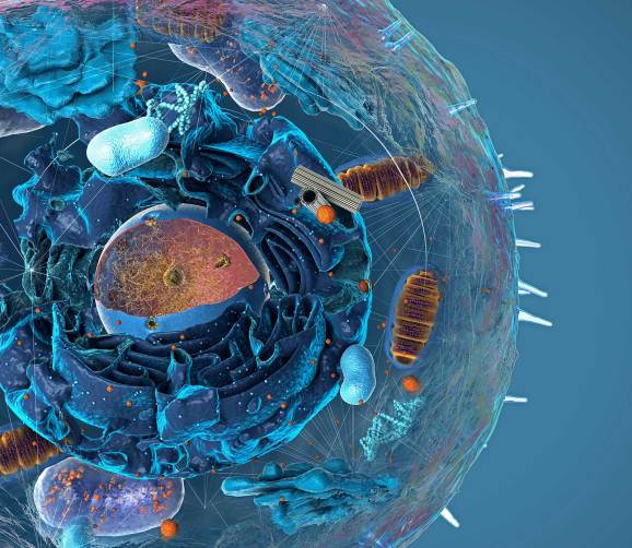
Visualize Cellular Phenotypes and Functional States in Context
Cellular detection is essential to spatial biology, revealing how individual cells behave, interact, and respond within their native tissue context. Enzo’s cellular detection solutions for spatial biology include robust assay kits designed to visualize and quantify key cellular processes—such as cell health, proliferation, and signaling—within complex tissue environments. Optimized for multiplexing and high-resolution imaging in both 2D and 3D models, these tools enable researchers to explore spatial relationships and cellular dynamics with precision and achieve high spatial and temporal resolution for detailed cellular analysis.
Comprehensive portfolio of cell-based assays and fluorescent dyes to monitor apoptosis, oxidative stress, cell proliferation, and mitochondrial health in 2D and 3D tissue models
Achieve high spatial and temporal resolution for detailed analysis of cellular processes within intact tissue architecture
Cited in nearly 3,000 peer-reviewed publications, supporting their broad utility across spatial biology, drug discovery, and disease modeling
Plaque Burden Detected with PROTEOSTAT® Aggresome Detection Kit
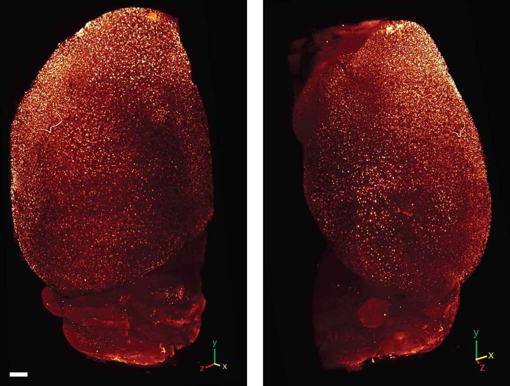
3D-reconstruction of MesoSPIM imaging of iDISCO-cleared and PROTEOSTAT® Aggresome Detection Kit (ENZ-51035) used to stain APP/SP1 mouse brain. PROTEOSTAT® dye staining allowed high-density data acquisition of plaque burden throughout a whole brain sample. Scale bar = 500 µm.
Spatial Relationship Between the Nucleus and Other Organelles
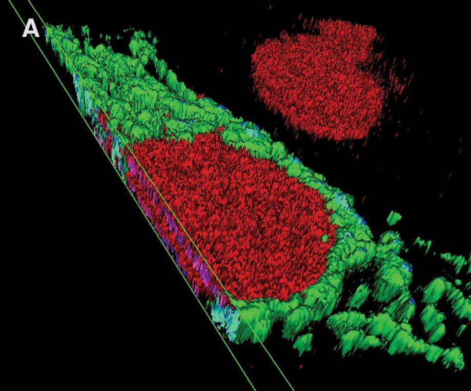
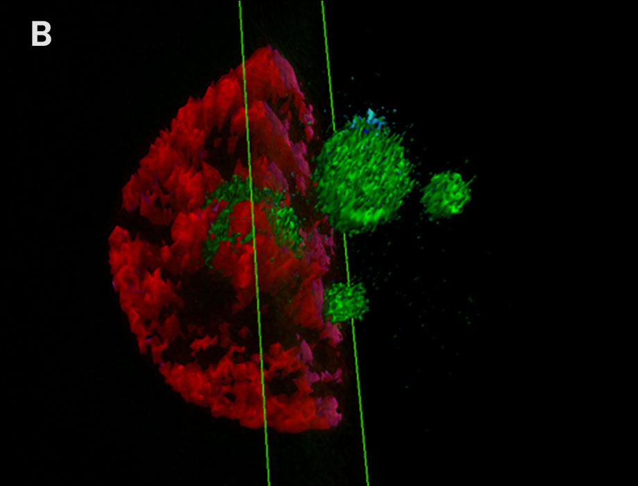
A. 3D-reconstruction of the spatial relationship between the nucleus (red) with NUCLEAR-ID® Red Cell Cycle Kit (ENZ-51008) and GFP-expressing mitochondria (green). B. 3D-reconstruction of the spatial relationship between the nucleus (red) and the nucleolus (green) with TOTAL-NUCLEAR-ID® Kit (ENZ-51006). Optical sections were imaged with conventional fluorescence microscope to create 3-D reconstruction.
Quantify Autophagic Vacuoles without Transfection with CYTO-ID® Autophagy Detection Kit
CYTO-ID® Autophagy Detection Kit 2.0 (ENZ-KIT175) was used to detect autophagy in HeLa cells cultured in (A) media under normal conditions, (B) starvation media (EBSS) treated with 40uM Chloroquine for 4 hours. Starved cells show a higher quantity of autophagic vacuoles compared to cells under normal conditions (fluorescent green).
Multiplex Hypoxia and Oxidative Stress Detection with ROS-ID® Hypoxia/Oxidative Stress Detection Kit
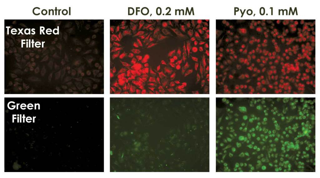
HeLa cells were seeded on microscope slides and treated the next day, with DFO (chemical inducer of hypoxia) or pyocyanin (oxidative stress inducer) for 4 h at 37°C as described in the manual. ROS-ID® Hypoxia/Oxidative Stress Detection Kit (ENZ-51042) was used to detect hypoxia (red) and oxidative stress (green). Post-treatment, slides were washed with PBS, cover-slipped and visualized using an Olympus BX-51 fluorescence microscope.
Featured CELLESTIAL® Cell-based Assays
Mitochondrial Detection
MITO-ID® Red Detection Kit (GFP-CERTIFIED®)
MITO-ID® Green Detection Kit
MITO-ID® Membrane Potential Cytotoxicity Kit
MITO-ID® Membrane Potential Detection Kit
Nuclei/Nucleoli, Cell Cycle, Transcription Arrest Analysis
NUCLEAR-ID® Red Cell Cycle Kit
NUCLEAR-ID® Green Cell Cycle Kit
Total NUCLEAR-ID® Green/Red Nucleolar/Nuclear Kit
NUCLEOLAR-ID® Green Detection Kit
NUCLEAR-ID® Green Chromatin Condensation Kit
Lysosomal and Autophagosome Detection
LYSO-ID® Red Detection Kit (GFP-CERTIFIED®)
LYSO-ID® Green Detection Kit
LYSO-ID® Red Cytotoxicity Kit (GFP-CERTIFIED®)
Endoplasmic Reticulum Detection
ER-ID® Green Assay Kit
ER-ID® Red Assay Kit (GFP-CERTIFIED®)
Whole Cell Analysis
GFP-CERTIFIED® Apoptosis/Necrosis Detection Kit
ORGANELLE-ID® RGB III Assay Kit