Lab Essentials
 AMPIVIEW® RNA probes
 Enabling Your Projects
  GMP Services
 Bulk Solutions
 Research Travel Grant
 Have You Published Using an Enzo Product?
Spatial Biology
Enabling Flexible Multiomics Analysis

From genomics to tissue analysis – combine any target, using any label, across any sample type
Our innovative and patented technologies enable flexible multiomics analysis, bridging the gap from fundamental genomics to intricate tissue architecture. As pioneers in labeling and detection technologies, Enzo was first to develop non-radioactive nucleic acid detection technologies, developing in situ hybridization probes for gene expression over four decades ago.
Our comprehensive and flexible reagents empower scientists to analyze any combination of targets – from genomics to tissue morphology – using any label and across any sample type. This flexibility supports both singleplex and multiples analysis, with manual or automated options for chromogenic or fluorescent detection. Accelerate your scientific discoveries and obtain deeper insights into biological processes, disease mechanisms, and therapeutic responses by leveraging the adaptability that surpasses the limitations of traditional singleomics approaches.
Whether using singleplex or multiplex assays, automated or manual protocols, and fluorescence or chromogenic detection, our flexible and adaptable solutions cater to diverse research needs

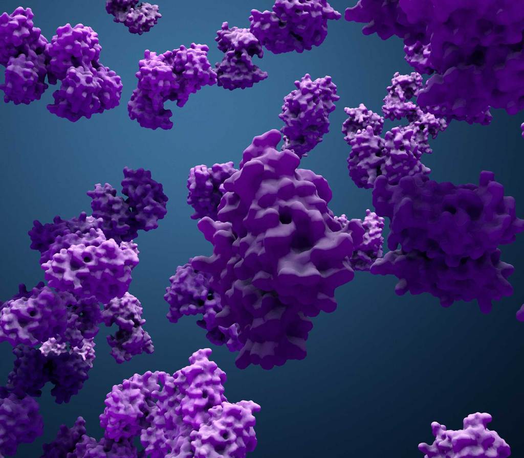
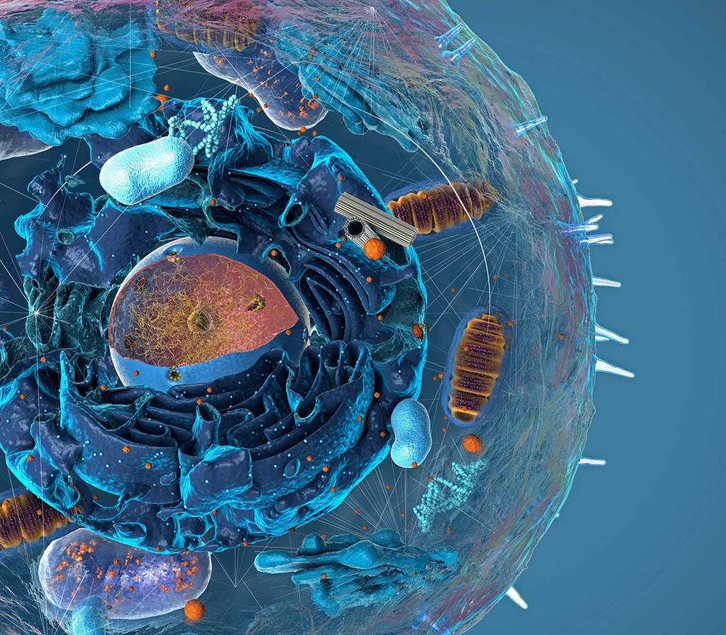
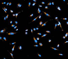
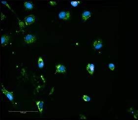
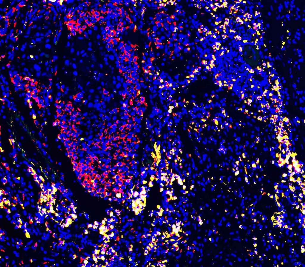
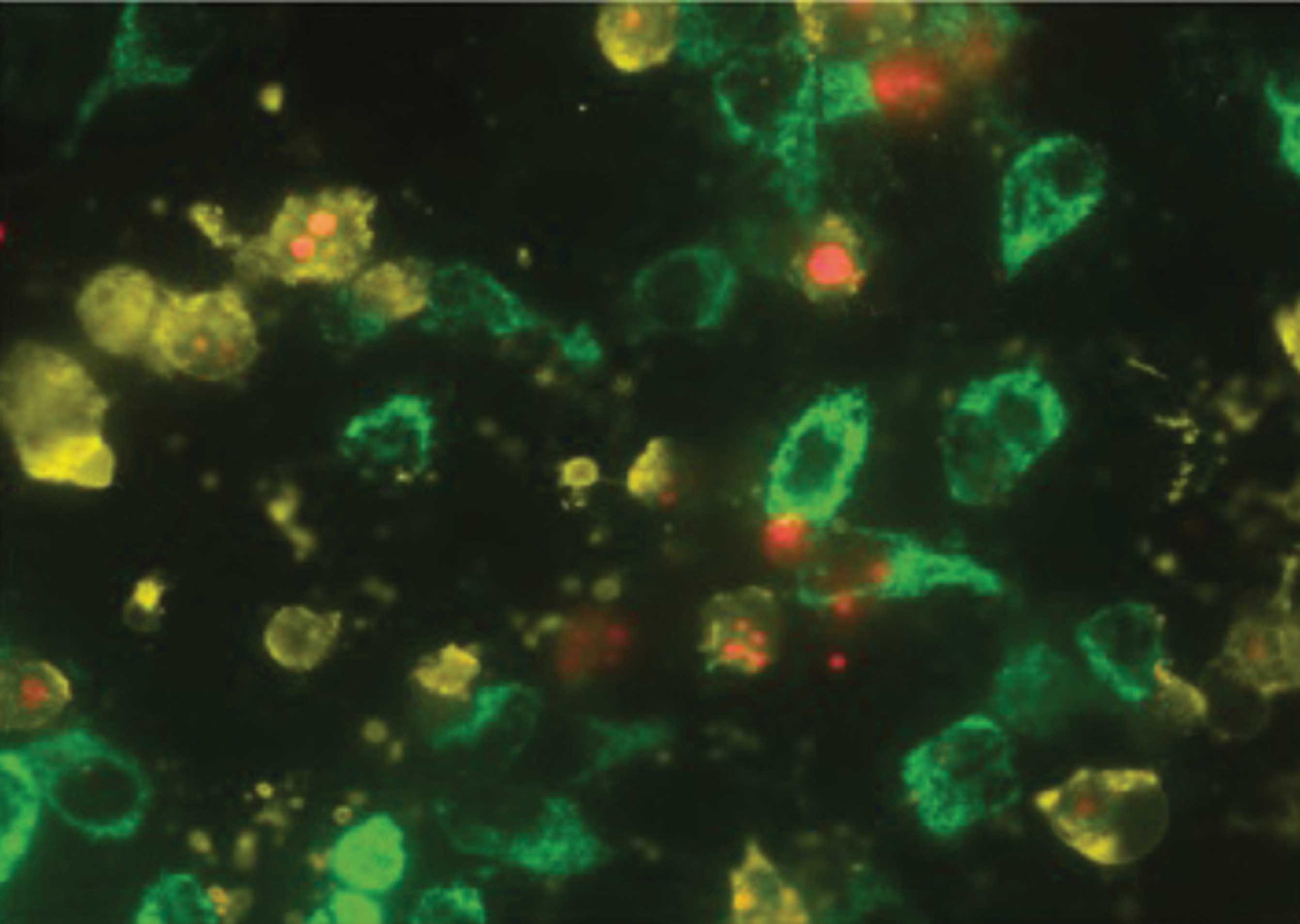
Simultaneously analyze multiple targets, including nucleic acids, proteins, and organelles.
Perform multiplexing individually or combine targets for comprehensive insights.

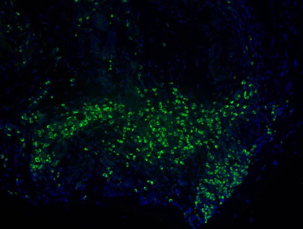
AMPIVIEW® RNA Probes
Powered by Enzo’s LoopRNA™ ISH Technology
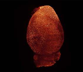
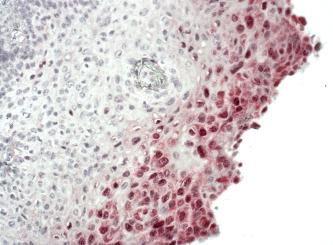
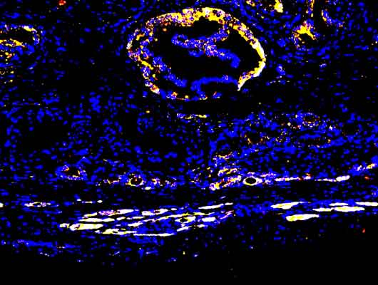
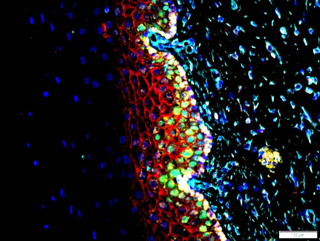
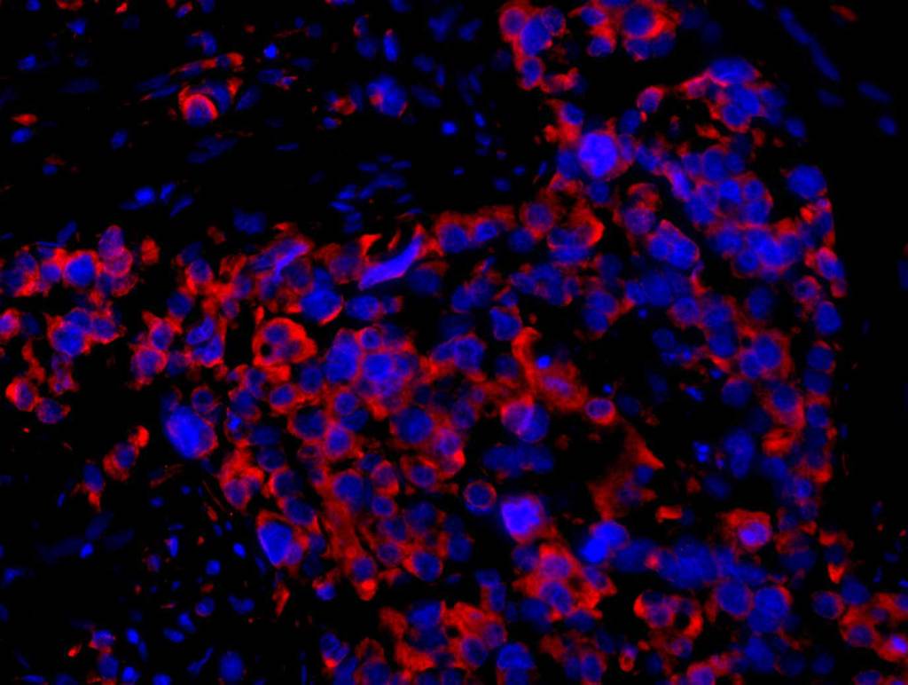
Enabling detailed spatial analysis, allowing researchers to visualize and understand the spatial context of gene expression and interactions within tissues.
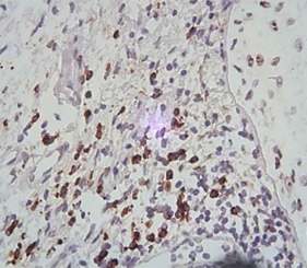
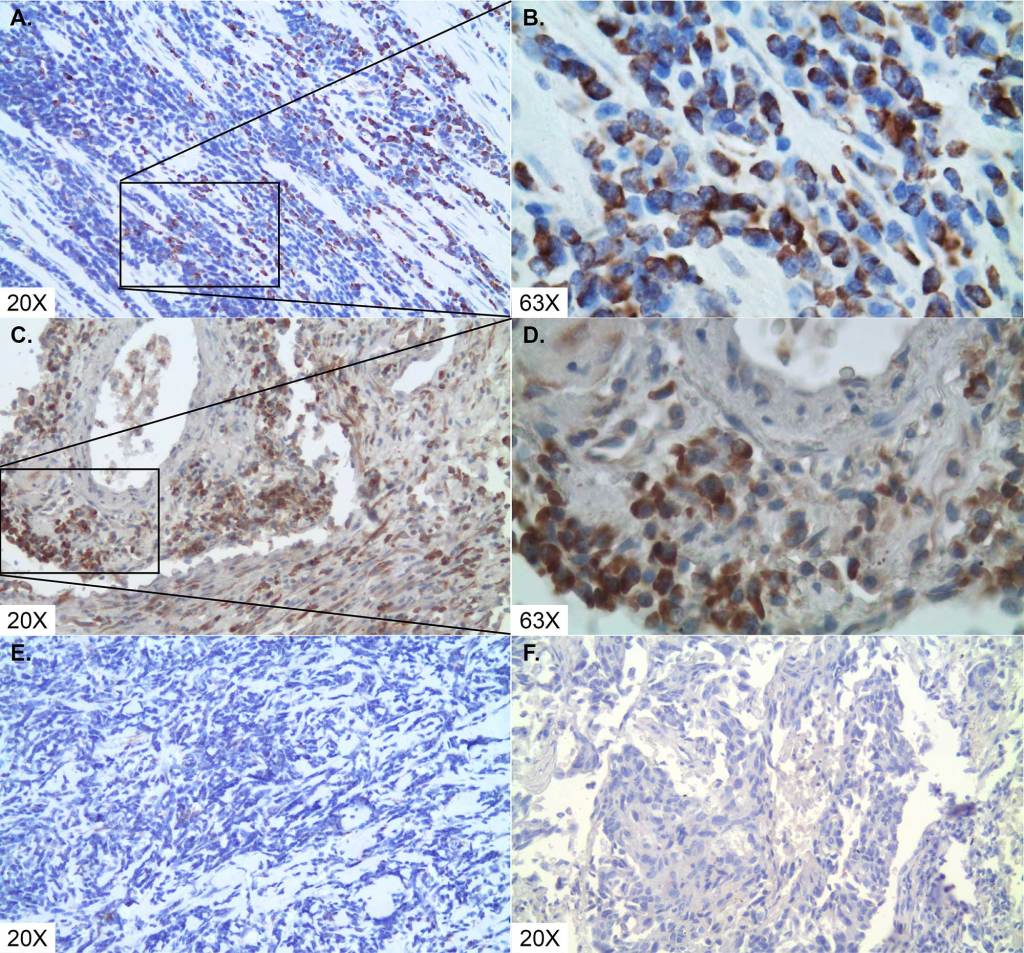
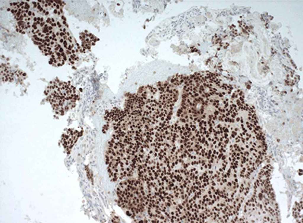
- High Sensitivity and Reliability: Superior sensitivity for detecting unique nucleic acid targets (DNA/RNA) down to a single cell level.
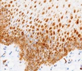
- Versatile Applications: Suitable for singleplex and multiplex assays in formalin-fixed, paraffin-embedded (FFPE) tissues and cells.
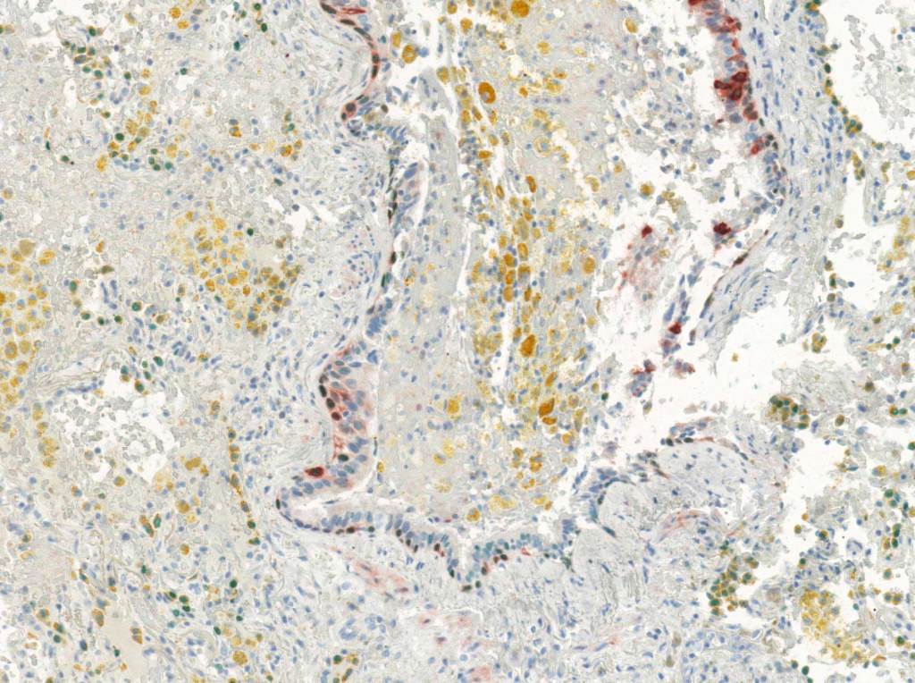
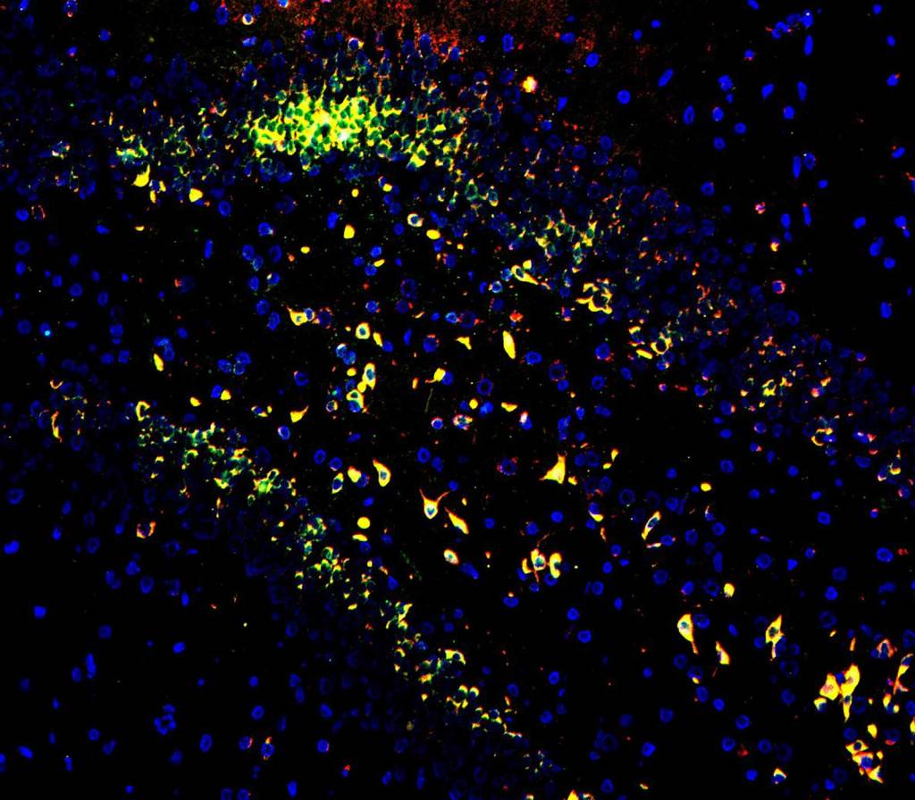
- Preserved Morphology: Mild protocols ensure that tissue morphology is maintained while providing clear visualization under the light microscope with chromogenic detection or fluorescence microscope with fluorescence detection.
- Flexible and Adaptable Workflows: Our open solutions are ready for both manual and automated workflows, ensuring compatibility with various research setups and IHC detection reagents.
- Customizable: Probes can be labeled with biotin, Dig, DNP, or FITC, enabling precise targeting of any gene across any species.
- Cost-Effective: Optimized with Enzo’s cost-effective IHC/ISH detection portfolio, making them a budget-friendly option for researchers.
Enzo’s custom services provide tailored solutions that empower spatial biology research from discovery to validation. With deep expertise in labeling and detection technologies, our team delivers custom services to support nucleic acid detection, protein localization and cellular analysis.
We offer specialized services to address the evolving needs of spatial biology applications, including:
- Custom AMPIVIEW® RNA probe design and production
- IHC, IF, and ISH staining services on FFPE or frozen tissues and cells
- Validation of AMPIVIEW® RNA probes for target specificity and tissue compatibility
- Antibody conjugation services (fluorophores, enzymes, biotin, haptens)
- Singleplex and multiplexing capabilites for RNA + protein + cellular co-detection
Enzo provides the technical depth and operational flexibility to accelerate your spatial biology programs.

With Enzo’s products, researchers can delve into the intricate spatial relationships within biological systems. Our products can perform both singleplex and multiplex assays, which have been validated by our own scientists, in addition to collaborators, beta testers and customers.
Webinar
Quantifying Targeted mRNA in Single Cells via In Situ Hybridization with Enzo AMPIVIEW®RNA Probes
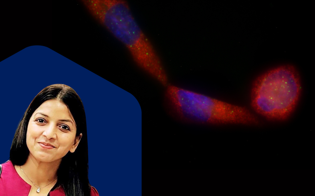
Webinar
Detecting Non-Coding RNAs with AMPIVIEW® ISH Probes

Latest Articles