Lab Essentials
 AMPIVIEW® RNA probes
 Enabling Your Projects
  GMP Services
 Bulk Solutions
 Research Travel Grant
 Have You Published Using an Enzo Product?
Francesca Catto, Ph.D. | Co-Founder and CEO | IMAI
Hartmut B.F. Pohl, Ph.D. | Application Scientist | Enzo Life Sciences
INTRODUCTION
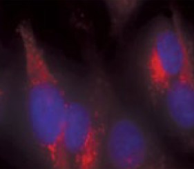
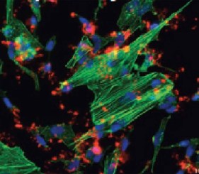
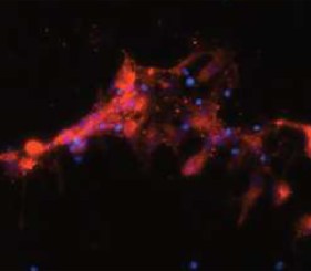
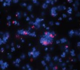
Protein aggregation and plaque formation are key hallmarks of several neurodegenerative diseases, including Alzheimer’s disease (AD), Parkinson’s disease (PD), Huntington’s disease (HD), amyotrophic lateral sclerosis (ALS), and prion diseases. Protein aggregation in these diseases ranges from random misfolded aggregates to inclusion bodies, and large extracellular deposits of highly compacted amyloid fibers of densely packed proteins in β-sheet conformation termed plaques. While plauques are not necessarily directly responsible for all aspects of neurodegeneration in the respective disease (impaired neuronal and glial cell metabolism, viability, or cellular atrophy) which ultimately cumulate in loss of neural function, plaque size, and density generally correlate well with the severity of the disease. Additionally, protein aggregates and amyloid plaques themselves can be disruptive to neuronal function and exhibit cytotoxicity. Plaque burden can therefore be both a valuable indicator of severity in these neurodegenerative diseases and a readout of intervention strategies.
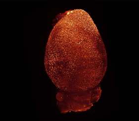
However, plaque size and density do not only show high degrees of heterogeneity throughout various central nervous system (CNS) tissues, but have also been demonstrated to show high variability in response to treatments targeting plaque burden throughout the CNS. How exactly plaque burden heterogeneity throughout CNS regions occurs, or why a response to treatment often shows high regional variability, both in lightening plague burden and recuperating neural function, remains largely unclear. The ability to easily image and quantify plaque burden throughout the CNS tissue and monitor regional and temporal heterogeneity is therefore crucial for disease assessment or analysis of treatment efficacy. Here, we describe whole organ plaque burden visualization through high-resolution light-sheet imaging of brains cleared with organic solvents and stained with Enzo’s PROTEOSTAT® dye.
Download this Application Note
Benefits
- Visualization of aggregates within an entire organ
- High density data on plaque burden
- Easy and fast protocol
The ease of use of the PROTEOSTAT® dye in combination with its robustness has made it easy to implement into IMAI’s proprietary tissue clearing and staining technology. The rapid results and high density data that can be obtained by staining 3D brain tissue samples with PROTEOSTAT® allow for the precise and large-scale analysis of protein aggregates plaguing a variety of neurodegenerative diseases.
Dr. Francesca Catto – CEO & Co-Founder – IMAI MedTech
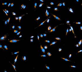
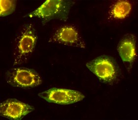
PROTEOSTAT® Aggresome detection kit
ENZ-51035
Robust, quantitative detection of aggresomes relevant to the study of neurodegenerative disease, liver disease and toxicology
| Application | Flow Cytometry, Fluorescence microscopy, Fluorescent detection |
|---|
Latest Articles