Lab Essentials
 AMPIVIEW® RNA probes
 Enabling Your Projects
  GMP Services
 Bulk Solutions
 Research Travel Grant
 Have You Published Using an Enzo Product?
Sarah Beckman, PhD1; Nadia A. Rana, PhD2
1 BioTek Instruments, Inc., Winooski, Vermont, USA
2 Enzo Life Sciences, Farmingdale, New York, USA
INTRODUCTION
The mitochondrion is a highly dynamic organelle, often called the “powerhouse of the cell” for its ability to produce cellular energy in an efficient manner. Mitochondria are the primary manufacturers of ATP, but they also regulate iron homeostasis and the production of free radicals. Mitochondria have a duality of function in that they are involved in the maintenance of viability and vitality, but also play a role in the regulation of apoptotic cell death. Studies have demonstrated that metabolic control through mitochondria is not only related to cell fate, but also plays an important role in differentiation. These diverse functions of mitochondria are all at some point dependent on the mitochondrial membrane potential (MMP).
Mitochondrial respiration generates an electrochemical gradient of protons made up mostly of a negative electrical potential difference across the mitochondrial inner membrane. During mitochondrial oxidative phosphorylation, the transfer of electrons through electron transport chain (ETC) complexes I-IV in the inner mitochondrial membrane, which provides the energy to drive protons against their concentration gradient across the inner mitochondrial membrane (out of the mitochondrial cytoplasm). The result of this process is an accumulation of H+ outside the membrane, which then flow back into the mitochondria through Complex V, thus producing ATP. This accumulation of H+ results in an electrochemical gradient, otherwise known as MMP.
Download this Application Note
Mitochondria are present in most of the cells in a living organism, and as such, they are implicated in a wide variety of diseases. Defects in the transfer of electrons across the mitochondrial membrane can cause electrons to accumulate on the ETC complexes and enhance reactive oxygen species (ROS) production. This accumulation increases the potential for electrons to bind with free oxygen species and contributes to many pathological conditions including degenerative diseases, cancer, and aging. Disruption of MMP is one of the earliest intracellular events to occur following induction of apoptosis. In mammalian cells, three responses of mitochondria following a death signal have been noted: a transient hyperpolarization of MMP, a subsequent substantial depolarization of MMP, and, in selected settings, the release of cytochrome c.
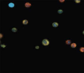
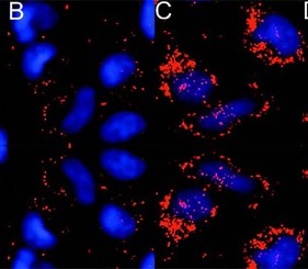
Thus, cell-based assays for analysis of MMP are extremely valuable in order to obtain insights into both cell disease and viability. Evaluating the functional status of mitochondria is critical to elucidating the role of mitochondrial activity in drug-induced toxicity, apoptosis, stem cells, and other cellular and biochemical processes. Quantitative microscopy of the intracellular distribution of membrane-permeant cationic fluorophores provides a means to measure MMP in live cultured cells. To assess the effects of carbonyl cyanide 3-chlorophenylhydrazone (CCCP) and ethanol on MMP, such a fluorescent probe in combination with object-based spot counting analysis. To accurately and efficiently determine the number of MITO-ID Membrane Potential positive aggregates per cell analysis was performed using the Gen5™ 3.03 software with object spot counting capability.
MITO-ID® Membrane potential detection kit
ENZ-51018
The only mitochondria membrane potential assay that monitors energetic status using a simple mix-and-read, no-wash protocol

| Application | Flow Cytometry, Fluorescence microscopy, Fluorescent detection, HTS |
|---|