Lab Essentials
 AMPIVIEW® RNA probes
 Enabling Your Projects
  GMP Services
 Bulk Solutions
 Research Travel Grant
 Have You Published Using an Enzo Product?
The proteasome is widely recognised as the central enzyme of non-lysosomal protein degradation. It is responsible for intracellular protein turnover and it is also critically involved in many regulatory processes and, in higher eukaryotes, in antigen processing. The 26S proteasome is the key enzyme of the ubiquitin/ATP-dependent pathway of protein degradation. The catalytic core of this unusually large (2000kDa, 450Å in length) complex is formed by the 20S proteasome, a barrel shaped structure shown by electron microscopy to comprise of four rings each containing seven subunits. Based on sequence similarity, all fourteen 20S proteasomal subunit sequences may be classified into two groups, α and β, each group having distinct structural and functional roles. The α-subunits comprise the outer rings and the β-subunits the inner rings of the 20S proteasome. Observations of the eukaryotic proteasome and analysis of subunit sequences indicate that each ring contains seven different subunits (α7β7β7α7) with a member of each sub-family represented in each particle. Each subunit is located in a unique position within the α- or β-rings. In addition to the 20S particle, the 26S complex contains over twenty additional proteins, ranging in molecular weight from 25 to 10kDa, located in a distinct complex called the ‘PA700 proteasome activator’ or the ‘19S complex’, and which determines substrate specificity and provides the multiple enzymatic functions necessary for proteolysis and viability. Systematic analysis of the sub-unit components have revealed at least six members to be ATPases belonging to a new family of ATPbinding proteins, together with a further fifteen sub-units that lack the capacity to bind ATP, isopeptidases and several other proteins thought to be responsible for the unfolding of a protein substrate prior to insertion into the proteolytic core of the 20S proteasome.
Shipping: Available products typically ship within 24/48h, via priority shipping.
Do you need support? Contact Customer Service or Technical Support.
Online Account
Access or Create Your Account
This antibody is covered by our Worry-Free Guarantee.

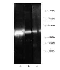
Western blot analysis: Composite luminograph of (a) HeLa S3 cytosolic preparation, (b) purified 26S proteasome, and (c) human placental proteasomefraction after SDS PAGE followed by blotting onto PVDF membrane and probing with antibody BML-PW8830. Antibody dilution 1:5000 using ECL procedure (1 min exposure).
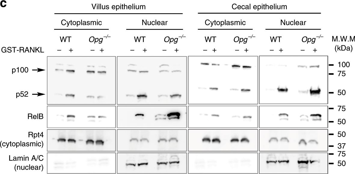
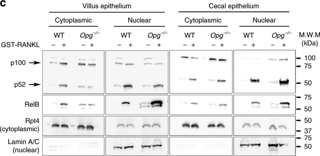
RANKL–RANK signaling is stimulated in the gut epithelia of Opg−/− mice.a GP2+ cells (red) are more effectively induced in the cecal epithelium (CE) and ileal villi (VE) of Opg−/− mice by RANKL administration. Whole-mount immunohistochemical images of Spi-B (green) and GP2 (red) in the VE and CE of WT (upper panels) and Opg−/− mice (lower panels) treated with either GST (control; left) or GST-RANKL (right). Scale bars: 100 µm. Representative images from three independent experiment are shown. b Quantitative PCR analysis of M-cell marker expression in conventional epithelia from the VE and CE of mice injected with GST (control) or GST-RANKL. Results were normalized to Gapdh expression and are presented relative to the expression in the ileal epithelium from GST-treated mice. Data shown are mean values from three independent experiments (error bars indicate standard deviation). ***p < 0.005, **p < 0.01, *p < 0.05; p values were calculated with the Student’s t-test (n = 3 biologically independent experiments). c, d Nuclear translocation activities of RelB and p52 following RANKL stimulation are enhanced in Opg−/− mice. c Western blot analysis of p100/p52 and RelB in the VE and CE of mice injected with GST or GST-RANKL. Rpt4, a subunit of the 26S proteasome, was used as an internal control for the cytoplasmic fraction. Lamin A/C (Lamin) was used as an internal control for the nuclear fraction. Data are representative of two independent experiments. d Right, single confocal planes of the cecal FAE from WT and Opg−/− mice. FAE monolayers were stained with anti-RelB (green) and anti-Spi-B (red) antibodies and with Hoechst 33342. Left, a bar graph summarizing the proportions of RelB-positive cells among total numbers of M cells (Spi-B-positive cells). ***p < 0.005; p values were calculated with the Student’s t-test (n = 4 biologically independent experiments). Scale bars: 20 µm. The source data underlying panels b and d and non-cropped scan images of western blotting (c) are provided as a Source Data file.
Image collected and cropped by CiteAb under a CC-BY license from the following publication: Osteoprotegerin-dependent M cell self-regulation balances gut infection and immunity. Nat Commun (2020)

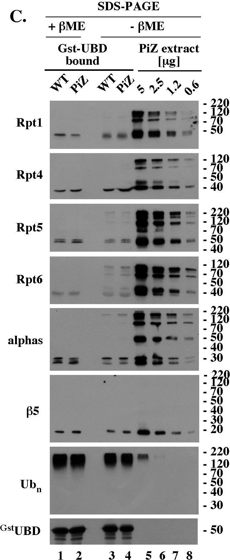
Reduction-sensitive modifications typical of aging WT mice accumulate prematurely on selected proteasomal subunits in the livers of PiZ mice.(A). Rpt4 Western blot analysis of unreduced, un-boiled liver extracts. 5 µg of the indicated extracts were mixed with Laemmli buffer without (unreduced samples) or with (reduced samples) βME (−/+ βME), separated by SDS-PAGE without prior boiling, and analyzed by Western blot with antibodies specific to Rpt4. (B). Rpt4 Western blot analysis of unreduced, but boiled, samples. Experiment like in A, lanes 1–9, except that extracts were mixed with Laemmli buffer without βME (- βME) and boiled for 4 minutes prior to SDS-PAGE. (C). Analysis of 26S proteasomes co-purified with polyubiquitin conjugates. WT and PiZ 26S proteasomes were co-purified with polyubiquitin conjugates as described in Fig. 4B and analyzed by Western blot after separation by SDS-PAGE with (lanes 1, 2) and without (lanes 3, 4) prior reduction by βME. Serial dilutions of unreduced liver extract from 103 old PiZ mouse are shown as reference (lanes 5–8). Extract prepared from 103 days old WT mice had similar reduction-sensitive modifications (data not shown, see panel A). (D). HPLC of WT and PiZ liver extracts followed by unreduced SDS-PAGE/Western blot analysis. Experiment like Fig. 5B, except that gel filtration fractions were not reduced with βME before SDS-PAGE. Data shown in A-D are representative of at least 3 independent experiments.
Image collected and cropped by CiteAb under a CC-BY license from the following publication: PiZ mouse liver accumulates polyubiquitin conjugates that associate with catalytically active 26S proteasomes. PLoS One (2014)
| Regulatory Status |
RUO – Research Use Only |
|---|
Related Products

| Application | ICC, IHC, WB |
|---|---|
| Host | Rabbit |
| Species Reactivity | Human |
| Alternative Name | Activating transcription factor 6 |
|---|---|
| Application | ChIP, ICC, IHC (PS), IP, WB |
| Host | Mouse |
| Isotype | IgG1 |
| Species Reactivity | Hamster, Human, Mouse, Rabbit, Rat |
| Alternative Name | p28 BAP31, B cell receptor-associated protein BAP31, BCR-associated protein BAP31, CDM, DXS1357E, 6C6-AG tumor-associated antigen, BCAP31 |
|---|---|
| Application | ELISA, Flow Cytometry, ICC, IHC (FS), IP, WB |
| Host | Mouse |
| Isotype | IgG2a |
| Species Reactivity | Human, Monkey |
| Alternative Name | Blm3, PA200 homolog |
|---|---|
| Application | WB |
| Host | Rabbit |
| Species Reactivity | Saccharomyces cerevisiae |
Last modified: May 29, 2024