Lab Essentials
 AMPIVIEW® RNA probes
 Enabling Your Projects
  GMP Services
 Bulk Solutions
 Research Travel Grant
 Have You Published Using an Enzo Product?
Western Blot
Enhanced Protein Detection

Simplify Your Western Blot: Six Simple Steps
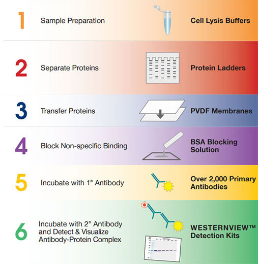
Western blotting is viewed as the gold standard for protein detection in molecular biology research. It is used to identify proteins within a cell or tissue lysate. Antibodies against your protein(s) of interest, bind to specific epitopes to identify the target protein within a lysate. Due to the high specificity of the binding, multiple target proteins can be identified on one membrane. Secondary antibodies then bind to your primary antibodies and when exposed to a substrate react, allowing for the visualization of the corresponding protein band.
Who wants to spend all that time in the dark?
We have removed the need for you to spend time in the dark room.
Let your blot develop before your eyes, right there on your bench!
The WESTERNVIEW® Detection Kits for Western Blot analysis quickly and clearly reveal bands on the transfer membrane without the need for specialized equipment.
Eventhough chemiluminescent detection is a common method of visualization this involves spending time in a tight dark room, with a timer and a list of exposure times, which are mostly chosen based on educated guesswork.
- A simple solution for real-time results – no film developer, fluorescent reader nor time in the dark room required.
- High sensitivity – quickly visualize your bands with minimal overexposure risk and with no visible background.
- Convenient and easy to use – includes secondary antibody and all reagents necessary to visualize your bands.
For high sensitivity, ease of use, and long-lasting signal, try our new WESTERNVIEW® Detection Kits
| Product | Kit Content |
|---|---|
| WESTERNVIEW® Detection Kit (Anti-Mouse) | AP Anti-Mouse Secondary, NBT/BCIP, Antibody Diluent, 10x Wash Buffer |
| WESTERNVIEW® Detection Kit (Anti-Rabbit) | AP Anti-Rabbit Secondary, NBT/BCIP, Antibody Diluent, 10x Wash Buffer |
| WESTERNVIEW® Dual Detection Kit (Anti-Mouse / Anti-Rabbit) | HRP Anti-Mouse Secondary, AP Anti-Rabbit Secondary, NBT/BCIP, DAB Chromogen, DAB Substrate Buffer, Antibody Diluent, SignaSure® Butter Salts, Tween®-20 |
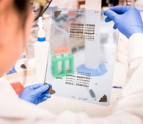
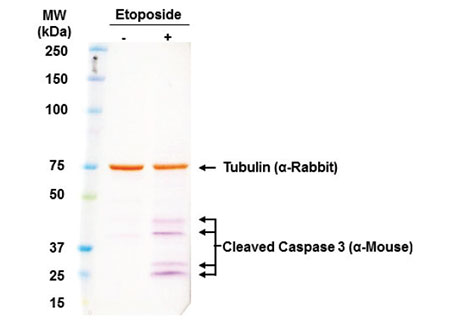
Reliably Distinguish Multiple Targets
PANC-1 cells treated with or without etoposide. Loaded total cell lysate (20 μg) and simultaneously incubated with tubulin and caspase antibodies before being developed with the WESTERNVIEW® Dual Detection Kit (Anti-Mouse / Anti-Rabbit).
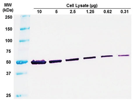
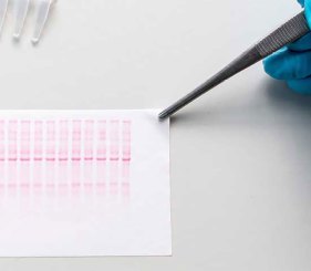
Sensitive Results Without Expensive Equipment
Total cell lysate from Jurkat cells. Membrane was probed with anti-tubulin. Detected using WESTERNVIEW® Detection Kit (Anti-Mouse) and exposed for 1 minute.
Clear Signals Without the Background
Jurkat cells treated with or without 12.5 μM etoposide for 18 hours. Loaded 12 μg lysate per well.

Do you want to know more about Western Blot?
Western Blot E-book
We have over 1,800 antibodies validated for Western Blot.
- Specific – to bind your protein of interest or primary antibody
- Sensitive – for high detection and low background
- Reliable – for consistent results
- Validated – for Western blot application and stated species
WB Antibody Search
Antibodies for Loading Controls
Find the right loading control to ensure proper interpretation of your Western blots. The sample type will help you determine which target to use for your loading control. Typically, loading controls are housekeeping proteins expressed at high and consistent levels.
Loading controls are used to:
- Check for equal transfer across membrane
- Ensure even loading between lanes
- Normalize and quantify your data
| Sample Type | Target | MW (kda) | PRODUCT NAME |
|---|---|---|---|
| Whole Cell & Cytoskeleton | Vinculin | 124 | Vinculin (human) monoclonal antibody (FB11) |
| Alpha Tubulin | 50 | α-Tubulin Recombinant monoclonal antibody (F2C) (Mouse IgG1λ) | |
| α-Tubulin Recombinant monoclonal antibody (F2C) (Rabbit IgGλ) | |||
| Beta Tubulin | 50 | β-Tubulin Recombinant monoclonal antibody (S11B) (Mouse IgG1λ) | |
| β-Tubulin Recombinant monoclonal antibody (S11B) (Rabbit IgGλ) | |||
| Actin | 42 | Actin polyclonal antibody | |
| Actin clonal antibody (S12-I) | |||
| Beta Actin | 42 | β-Actin polyclonal antibody | |
| GAPDH | 36 | GAPDH monoclonal antibody (GA1R) (Biotin conjugate) | |
| Mitochondrial | HSP60 | 60 | HSP60 monoclonal antibody (LK-1) |
| HSP60 monoclonal antibody (LK-2) | |||
| HSP60 monoclonal antibody (Mab11-13) | |||
| HSP60 polyclonal antibody | |||
| VDAC1 | 31 | Voltage-dependent anion channel polyclonal antibody | |
| Nuclear | Lamin B1 | 66 | Laminin B chain (human) monoclonal antibody (DG10) |
| HDAC1 | 5 | HDAC1 polyclonal antibody | |
| PCNA | 29 | PCNA monoclonal antibody (SPM350) | |
| Serum | Transferrin | 77 | Transferrin polyclonal antibody |

Enzo Antibodies
Sensitive. Specific. Consistent.
And now, On Sale!
* Antibody order must be placed on website to qualify for discount. See terms and conditions for offer details
Optimized for Western Transfer – Polyvinylidene Fluoride (PVDF) Membranes
- Sensitive protein detection with low background and very low burn-through
- Provides high surface area for strong hydrophobic interactions (adsorb 50% more protein than nylon or nitrocellulose.)
- Broad solvent compatibility
Membrane Specifications
| Protein Binding Capacity | 100-300 μg/cm2 |
| Solvent Resistant | Resistant to acetone, DMSO, dimethyl formahide, methanol, trifluoroacetic acid, and triethylamine. |
| Binding Interaction | Hydrophobic |
| Pore Size | 0.2 μM |
| Total Protein Stain Compatibility | Amido black, Ponceau S, Colloidal gold, Colloidal silver, India ink, Coomassie blue |
| Double-blotting Method | Yes |
| Strip and Re-probe | Yes |
| Detection Methods | Chromogenic, Chemiluminescent, Fluorescent, Radioactive, Chemifluorescent |
| Other Applications | Amino Acid Analysis, Protein Sequencing, Solid Phase Assay Systems |
| Available Formats | PVDF Transfer Membrane (7 x 9 cm) PVDF Transfer Membrane (10 X 15 cm) |
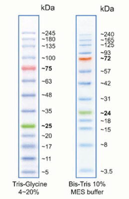
Protein ladder for use with protein gel electrophoresis
- 3 μL or 5 μL per loading for clear visualization during electrophoresis on 15-well or 10-well mini-gel, respectively
- Three color protein standard with 13 prestained proteins
- Apply more for thicker (>1.5 mm) or larger gel
Our protein ladder is designed for monitoring protein separation during SDS-polyacrylamide (PA) gel electrophoresis, verification of Western transfer efficiency on membranes (PVDF, nylon, or nitrocellulose) and for approximating the size of proteins. The ladder is supplied in gel loading buffer and is ready to use. Do not heat, dilute, or add reducing agent before loading.
Proteins are covalently coupled with a blue chromophore except for two reference bands (one green and one red band at 25 kDa and 75 kDa respectively) when separated on SDS-PAGE (Trisglycine buffer).
Assay and Validation Testing Services
We test, so you can discover.